Rotator Cuff Tear Treatment

Read on and find out everything you need to know about the rotator cuff treatment!

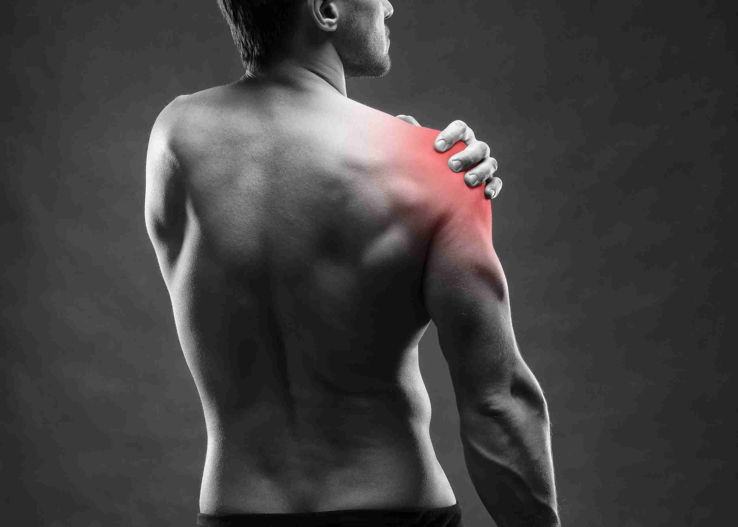
Causes and Risk Factors of Rotator Cuff Tear

A primary cause of tear of the rotator cuff is the natural wear and tear of tendons with age. This wear occurs from the age of 45. Statistics show that the rupture of the shoulder tendon affects 20% of people over 65 and 50% of people over 80. In addition, several factors can aggravate the natural wear of this cap, namely:

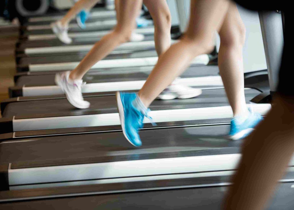
  • the current practice of a specific activity requiring a strong solicitation of the shoulder, such as in sports and other repeated activities, as mentioned above
  • smoking (resulting in the clogging of the arteries supposed to irrigate the muscles of the shoulder)
  • diseases such as diabetes
  • anatomy of the shoulder favoring natural wear

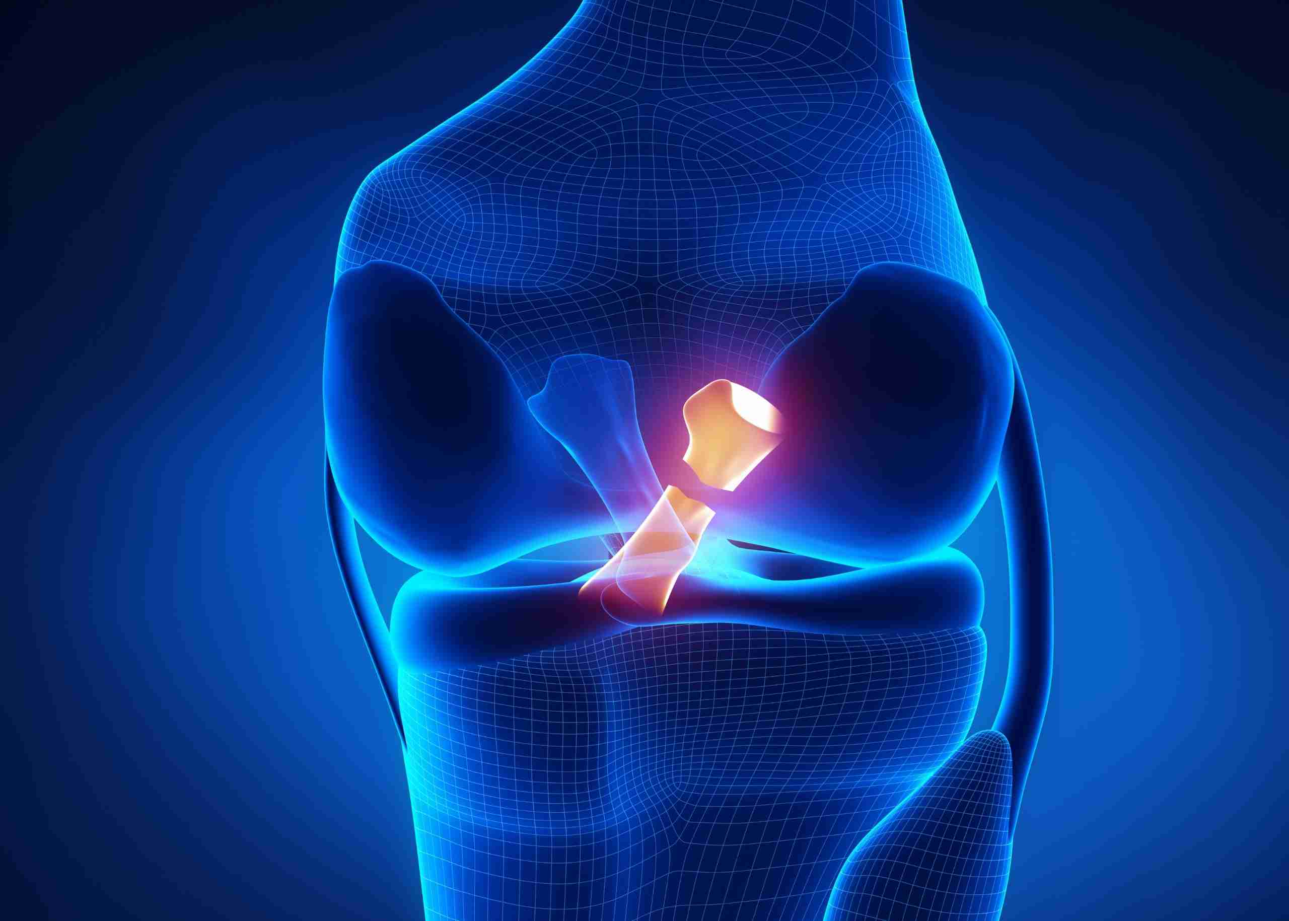
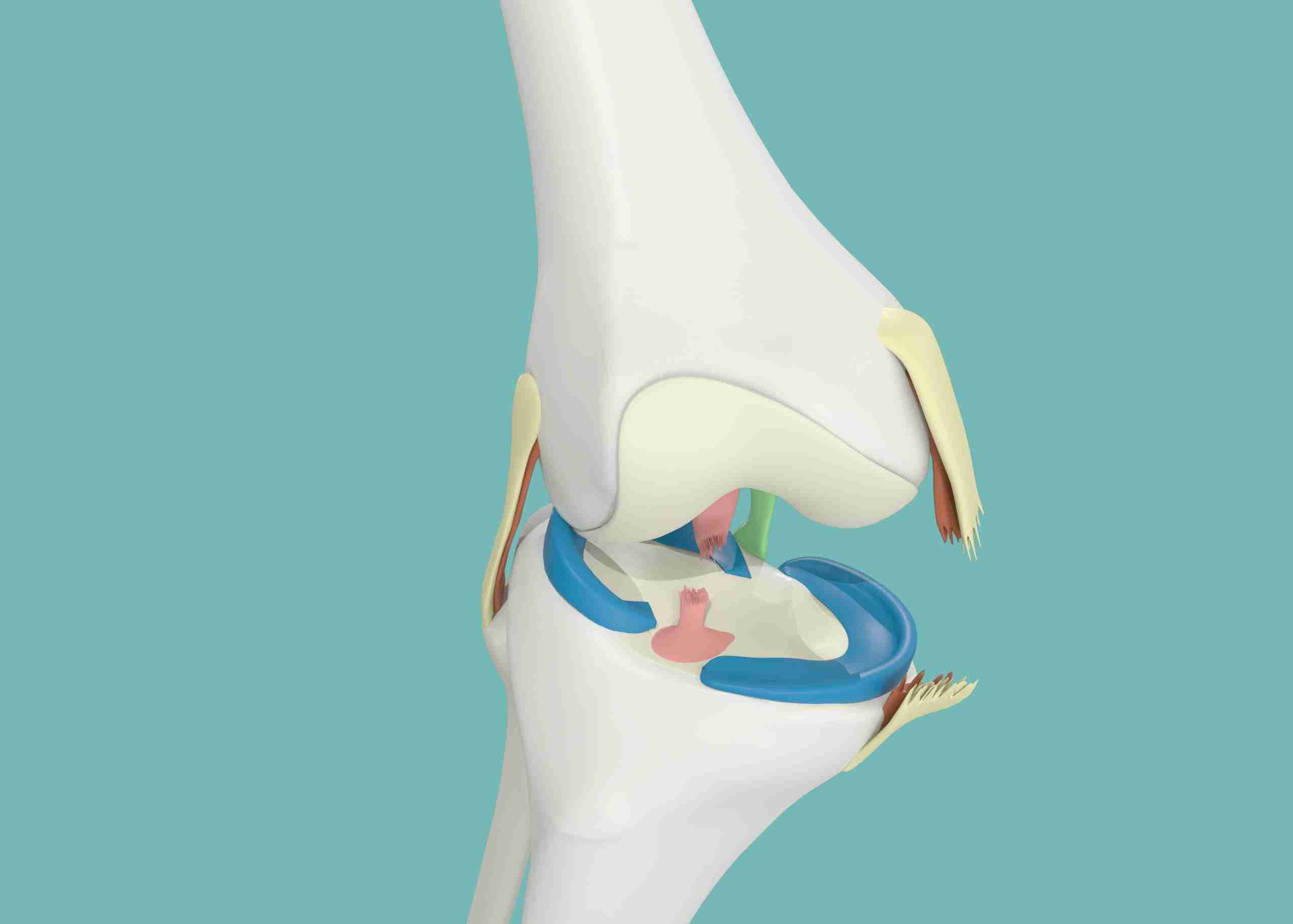
The rupture is the last phase of the subacromial syndrome. It happens when the space between the humerus head and the acromion is narrowing. The rotator cuff tendons (mainly the supraspinatus tendon) are located in this space, protected from the acromion bone by a bag. This bag is known as the subacromial bursa.

The decrease in this space has many causes. The most frequent one is the anatomical constitution of your shoulder (hook-shaped acromion, calcification of the Cossack-acromial ligament, etc.). If we add the repeated movement along the years, it produces a progressive rupture due to the collision between the supraspinatus and the acromion. On the other hand, unlike other tendons in the body, this tendon has very little self-repair capacity in the face of degenerative changes.

In addition to this slow rupture mechanism, there are also traumatic ruptures, in which the tendon ruptures after a fall with an outstretched arm: acute supraspinatus rupture. Sometimes, the rupture of the rotator cuff occurs following a simple and innocuous gesture or a false movement (more common in people over 50). This is called trophic rupture of the shoulder tendon and can also happen to athletes and sports performers.

Rotator Cuff Tear Diagnosis

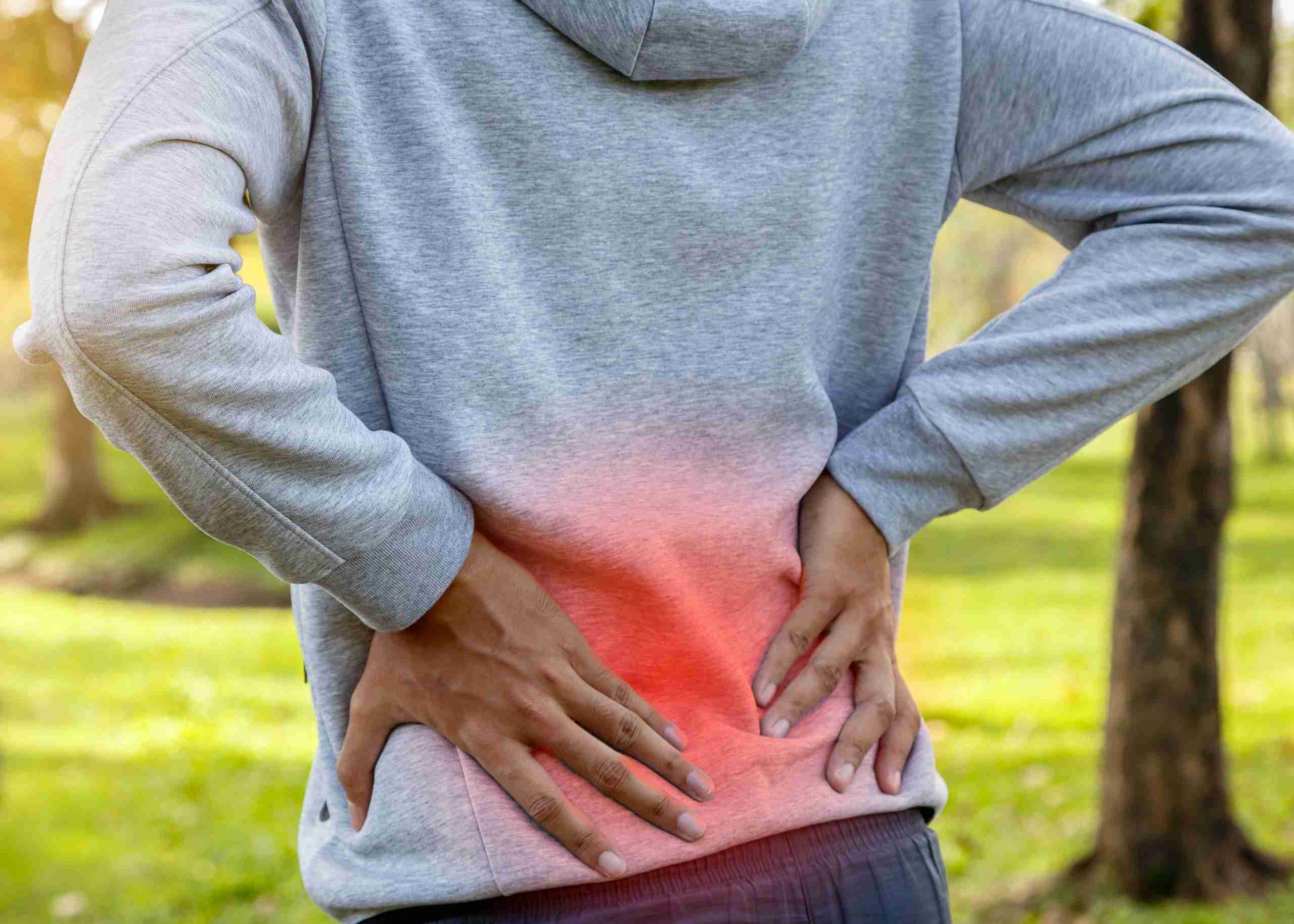
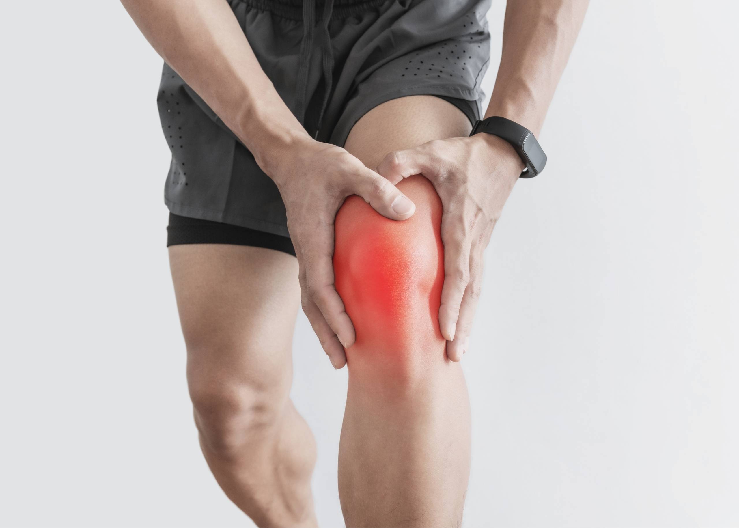
The diagnosis of complete rupture of the supraspinatus tendon is initially clinical and with imaging tests. The person who presents a rotator cuff tear complains of pain in the shoulder, especially with mobilizations and often pain at night or the inability to lift the arm correctly.

The main diagnosis of tear of the rotator cuff is through a shoulder MRI. The MRI shows us the rupture of the tendon. It also gives us information about the gap or space of the rupture and if there is atrophy or loss of musculature. This also informs about the condition of other tendons, such as the subscapularis or infraspinatus.

Ultrasound is another diagnostic test that, in good hands, allows good visualization of the tendon and the rupture of the supraspinatus. ​Through it, we can obtain other fundamental data, including:

  • Affected tendon(s): Usually supraspinatus or supraspinatus accompanied by infraspinatus or subscapularis
  • State of the tissue and the tear: This information is essential to decide the treatment and if the tear is “repairable.”
  • Involvement of other structures such as articular cartilage and the long tendon of the biceps

With all this information plus the time of evolution and the symptoms, the doctor will propose a conservative treatment of the rupture (without repairing it) or a surgical repair by arthroscopy to the patient.

Rotator Cuff Tear Treatment

Usually, it is recommended to carry out a sequential treatment in 3 phases.

  • Phase 1 treatment with rehabilitation or physiotherapy
  • Phase 2 treatment with infiltrations
  • Phase 3 treatment with surgery

Non-Surgical Treatment

The aim is to recover the shoulder function (mobility and strength) as much as possible, dispensing with the ruptured tendon. The tendon will be broken, but the Orthopedic doctor will try to increase the rest of the muscles in the area to get closer to the state we had before the muscle broke.

It is recommended to start with a rehabilitation/physiotherapy treatment. There are many exercises and guidelines to recover after a partial tear in the rotator cuff. It is important to note that the exercises must be performed daily for the treatment to have an effect. Usually, it is a 6-12 week program performing cuff strengthening exercises five days a week.

The objective of the initial guideline through exercises or recovery is to achieve the correct shoulder mobility and strengthen the muscles surrounding the shoulder to achieve muscular stabilization of the shoulder. One of the causes of the rupture is the lack of synchronization or correct functioning of the articulation.

Analgesics are used for the initial pain, including corticosteroid injections, to reduce inflammation and pain. If physiotherapy has not effectively improved pain, the doctor can recommend the infiltration of hyaluronic acid.

Surgical Treatment

Shoulder arthroscopy is done in cases where it is decided to perform a rotator cuff tear repair due to the patient’s characteristics (active and in good general condition) and the tear (good tissue, little muscle shortening).

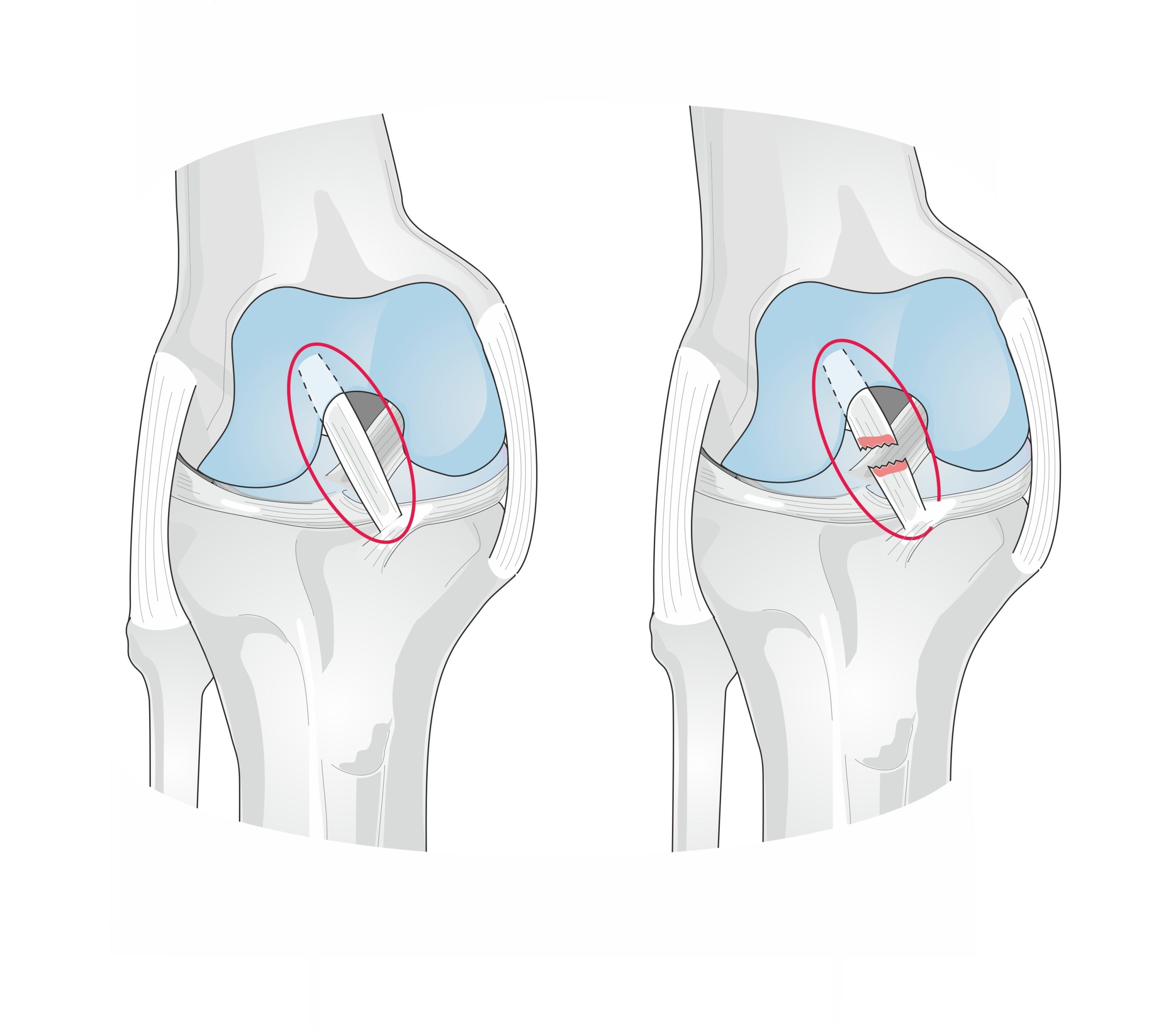
Shoulder arthroscopy is performed, and the tendon rupture is sutured using several anchors that join the tendon to the bone.

In this intervention, several surgical procedures are performed at the same time, all through 2 or 3 small incisions (less than 1cm)

  • Remove the inflamed bursa: Bursectomy
  • File the thickened acromion to increase the space in cases where it is: Acromioplasty
  • Repair the break: Cuff Suture

This repair is done by placing implants in the humerus (made of titanium or other biocompatible materials) to which strong tension-resistant sutures are attached. In cases of partial ruptures, it is often necessary to complete the tear and move the entire released tendon back to its original insertion site, which the surgeons join with one of the harpoons (implant).

In the case of total ruptures of one or two tendons, several implants are required to repair them.

At present, the new arthroscopic techniques and      anchoring systems achieve a very solid and stable fracture union, improving recovery time     , usually ranging from 2 to 4 months.

It is necessary to carry out an initial phase of rest that ranges between 2 and 4 weeks. Subsequently, physiotherapy is carried out to improve the shoulder joint’s mobility and musculature.

Irreparable Rotator Cuff Tear Treatment

When the rupture is massive, with stage 3 or 4 fatty degeneration and muscle atrophy, a latissimus dorsi flap in a motivated young patient is recommended, provided that the subscapularis tendon is present.

In a patient over 70, a biceps tenotomy is proposed after ineffective functional treatment. A biceps tenotomy is the cutting of the long biceps to reduce anterior pain under arthroscopy.

For other more complex forms, a modification of the shoulder functions by placing an inverted prosthesis is indicated. This prosthesis will use the upward forces of the deltoid muscle to transform them into rotational forces.

In case of a rotator cuff tear, the best is to consult experts with a distinguished history in complex surgical and nonsurgical procedures. The Orthopedic Surgeons team at the Burjeel Hospital for Advanced Surgery, Dubai, certified and Fellowship-trained, are the leading experts in performing complex shoulder surgeries with immense experience in treating all upper limb conditions due to arthritis or sports injuries. Call +971 800 55 or visit our website to book an appointment today!

Our Experts

See More

Spine Care (Back Pain) – Symptoms, Causes & Treatments

Read on to find the causes, symptoms and treatment of back pain!

Who doesn’t have back pain? Back pain refers to intense or moderate pain or a feeling of blockage in the spine. It often lasts for months, causing chronic back pain. There are sevral kinds of pathologies that  the spine, affecting every two out of five people, in which the most prevalent cases are of lower back pain. All age groups are affected depending on the pathology in question.

In the elderly, the back pain is often degenerative, related to age and the breakdown of the intervertebral discs, complicating daily life. A sedentary lifestyle accelerates this process. In the youngest, it is often due to malformative pathologies. And in working adults, it can be due to traumatic pathologies, and the repercussions of pain and incapacity can be major, as is often the case, compromising the personal and professional life. Statistics indicate that 50% of all working adults report back pain symptoms every year.

But back pain can have many other explanations. Therefore, it is essential to determine the cause of the back pain to direct the right treatment. In this article, we will try to explain the concepts related to back pain as clearly as possible to make it easier for you to understand your back problem.

Back Pain, the Most Common Issue

Back pain is widespread, according to statistics. Indeed, this worldwide scourge has already affected nine out of ten people. Let us add that a huge population of these individuals declare having suffered from their back more than ten times during the last five years. Also, more than half of the world’s population is regularly affected by back pain.

Finally, the working population is the most affected: more than four out of five active people report having back pain during or after their working hours, according to a survey.

There are two types of back pain:

  • Mechanical back pain: The pain is more intense during the day, during an activity.
  • Inflammatory back pain: The pain is more severe in the evening and at night and generally fades during the day after morning “strengthening.”

Causes of Back Pain

A distinction must be made between localized back pain, characterized by pain from the dorsal spine, and referred back pain, which is symptomatic of another pathology affecting a neighboring organ.

As a reminder, the dorsal column is the part of the vertebral column made up of the thoracic vertebrae. It is located between the neck (cervical vertebrae) and the lower back (lumbar vertebrae). The dorsal column consists of 12 thoracic vertebrae, which are fused to the ribs.

Localized Back Pain

The pain localized at the level of the dorsal column is not a question of pain projected from another organ. There are several underlying causes:

  • The degeneration of the intervertebral discs: It can cause osteoarthritis of the back or a herniated disc: over time, the intervertebral disc becomes less flexible. It loses its role as a shock absorber and can trigger acute or even chronic back pain. In case of severe deterioration, the nucleus pulposus (gelatinous substance located in the heart of the intervertebral disc, which acts as a shock absorber and ensures the vertebrae’s movement) can penetrate the spinal canal, causing a herniated disc whose volume is very variable. These pathologies can also be linked to wear and tear when the back is overworked.
  • Chronic rheumatic and inflammatory diseases: Arthritis, spondyloarthropathy (including ankylosing spondylitis), spondylodiscitis (infection of the intervertebral discs and adjacent vertebral bodies), etc., fall in this category.
  • The vertebral fracture: It can be of traumatic origin or linked to underlying osteoporosis (vertebral compression). Trauma occurs during a brutal shock, an accident or a fall. Note that the spine protects the spinal cord, and when the latter is affected, the consequences can be fatal or lead to paraplegia or serious neurological damage.
  • Prolonged poor posture: This is particularly the case for patients who work long hours in a seated position in front of their computer. Poor posture during sleep can also cause back or neck pain.
  • Pregnancy or obesity (especially visceral.)
  • Hormonal changes: Menstrual cycles, pregnancy or menopause in women.
  • An intraspinal tumor.
  • Bone deformity diseases.
  • Paget’s disease: chronic bone disease. It causes bone hypertrophy and weakening in certain localized regions.
  • Kyphoscoliosis: deformation of the vertebral column, which causes a lateral deviation (scoliosis) and a deviation with posterior convexity (kyphosis.)
  • Spinal growth dystrophies or osteochondrosis: alteration of the structure of the vertebral disc, which affects children and adolescents. It can cause growth disturbances. Scheuermann’s disease is one of these pathologies.
  • Psychological disorders: Depression and chronic stress can cause muscle tension and back pain.

Referred Back Pain

Referred back pain is symptomatic of an attack or a disease affecting another organ. It could be:

  • A digestive disease: esophagitis, gastric or duodenal ulcer, cancer of a digestive organ (pancreas, stomach, esophagus), pancreatitis, etc.
  • A lung or pleural condition: pleurisy, lung or bronchial tumor, etc.
  • A cardiovascular disease: pericarditis, thoracic aortic aneurysm, coronary insufficiency, etc.

Symptoms of Back Pain

The pain is localized at the back and, more precisely, between the neck and the waist. The pain can be acute or, on the contrary, chronic (when it lasts more than three months). It is sometimes localized and sometimes diffuse. It is more or less intense and can look like:

  • A feeling of electric shock, stabbing or burning;
  • Tingling;
  • Back blockage;
  • Breathing discomfort;
  • A strain.

In case of back pain, consult a doctor to eliminate any underlying pathology. If the symptoms present themselves intensely, especially after a shock or a false movement, contact an Orthopedic doctor or the emergency department. It must be understood that the decision to treat (medically or surgically) depends above all on the symptoms. The nature of the lesion guides the choice of the optimal spine care treatment.

Diagnosis

The clinical examination

  • It consists of questioning the patient about the pain (location, improvement depending on the position, evolution, response to anti-inflammatories and analgesics, underlying psychological disorder or chronic disease, etc.);
  • Examination of the spine (palpation, flexion and flexibility exercises, etc.);
  • Pulmonary examination (stethoscope, breathing exercises, etc.);
  • Cardiac examination (heart rate, stethoscope, blood pressure, questioning about possible discomfort or chest pain, etc.);
  • Digestive and hepatic examination (palpation, clinical examination looking for possible jaundice, ascites or bruising, questioning about possible digestive disorders, etc.);
  • Neurological examination (study of walking, standing, muscle strength/tone, reflexes, sensitivity, motor coordination, questioning the patient about any neurological symptoms, etc.).

Imaging Examination

Imaging tests, such as X-rays, ultrasounds, CT scans and MRIs, may be required to identify the cause of the back pain.

Complementary examinations

  • Blood tests (in particular looking for biological markers of inflammation);
  • Imaging of the thoracic spine (scintigraphy, scanner, MRI, etc.);
  • Gastric imaging (fibroscopy, endoscopy, colonoscopy, etc.);
  • Cardiovascular examinations;
  • A lung ultrasound;

Treatments of Back Pain

When back pain is a disease symptom, the underlying disease should be treated. However, pain relief requires:

  • Nonsteroidal anti-inflammatory drugs (NSAIDs) or painkillers
  • The practice of regular physical activity or sport to strengthen the back and the abdominals;
  • physiotherapy and osteopathy can be effective;
  • Psychological care in the event of an underlying anxiety-depressive disorder;
  • Change certain habits, such as posture and ergonomics at work, carrying heavy loads, etc.
  • Alternative medicine: mesotherapy (micro-injections of local action products), acupuncture, etc.
  • Rest can sometimes be recommended over a short period.

Bed rest, anti-inflammatories and rehabilitation (once the acute phase has passed) can relieve back pain but cannot treat spinal fractures. If you have been diagnosed with a vertebral fracture caused by any of the varied reasons discussed above, the doctors can consider several innovative treatment options.,

Surgical Treatment for Spine Fracture

Conservative Treatment

Traditional treatment for back pain and/or vertebral fractures may include several days of bed rest, painkillers and, less commonly, a lumbar corset. Conservative treatment can reduce pain, but it cannot repair the spine.

Vertebroplasty and Kyphoplasty

The second treatment offered is vertebroplasty, which involves injecting a resin (called cement) into the vertebra. This procedure is performed under radiological control through a mini-incision of less than one cm. By hardening, this resin will suppress the micro-movements of the fracture, thus limiting the pain. It allows consolidation in a good position and avoids the appearance of a secondary deformation but does not allow the existing deformation to be corrected.

For this, Kyphoplasty is considered an evolution of the technique of straightening the vertebra. The surgeon uses an instrument such as a mechanical expander or an inflatable balloon before injecting the surgical resin. The major interest of Kyphoplasty is based on better correction of the vertebral deformity caused by the fracture

Spinal Laminectomy

Spinal Laminectomy is a decompression Surgery performed under general anesthesia by the posterior route through an incision in the lower back. There are several types of decompression surgeries ne of which is Laminectomy.

A laminectomy is a spinal decompression surgery performed on the lower part of the spine. It consists of removing part of the bone of the vertebra, the lamina, to relieve the pressure exerted on the spinal cord or one, or even several, spinal nerves.

Discectomy

A herniated lumbar disc can be the cause of sciatica by compression of a nerve root. The procedure performed to relieve symptoms is called a Discectomy, in which the herniated disc is removed but not completely, and the disc is cleaned.

During the procedure, a small incision is made over the site of the herniated disc. The surgeon then inserts the tubular retractor, which moves the muscles apart without cutting them.

The ligament that lines the back of the spine is opened, providing a view of the nerve and disc. The surgeon carefully pushes back the nerve and removes the herniated disc.

Foraminotomy

We speak of recalibration when it is a gesture limited to one level (1 disc). These procedures include a foraminotomy, which involves widening the foramen (the bony channel through which the spinal cord nerves pass) to create a larger opening and relieve the pinched nerve.

Depending on the state of health, it can be done using an open or minimally invasive surgical technique. These gestures can be combined if there are several levels to be treated.

Nucleoplasty

Nucleoplasty is another procedure recommended for disc decompression. The procedure consists of puncturing the nucleus pulposus of the affected disc through the skin with a needle or electrode, reducing the bulging of the disc and thus freeing the compressed nerve.

We hope the information we shared has helped you understand back pain’s causes and treatment options. Of course, this information will never replace the explanations given in consultation but constitute additional help. Indeed, we believe that your adherence to the treatment will largely depend on the quality of your information.

In case of back pain, the best is to consult an Orthopedic Spine Surgeon with unparalleled expertise in treating varied spine conditions and injuries. The Orthopedic Spine Surgeons at the Burjeel Hospital for Advanced Surgery, Dubai, are board-certified and experienced in minimizing the effect of neurological conditions on children and adults by providing optimal care and making informed treatment decisions.

Our Experts

See More

Posterior Cruciate Ligament (PCL) Injury

PCL injury is common in sports that require high knee flexion and pivoting, such as soccer, basketball, skiing and American football. Most often, an athlete will hear or feel popping in their knee as the injury occurs.

What is the Posterior Cruciate Ligament (PCL) 

The Posterior Cruciate Ligament (PCL) is one of the four major ligaments that connect your knee bones. It extends from the back of the tibia to the back of the femur, connecting them. 

The PCL helps prevent hyperextension of your knee, which is when your leg bends backward past its normal range of motion. The PCL helps stabilize and protect your knee joint when you pivot, cut or change direction. 

If you tear or rupture the PCL, it may not heal on its own. If this happens, you may need surgery to repair or replace your ligament with a tendon or cadaver tissue graft. 

Causes of PCL Injury 

Here are some common causes of posterior cruciate ligament injuries: 

  • Anterior knee pain—The anterior cruciate ligament is the most commonly injured knee. It can be caused by a blow to the front of the knee or by excessive flexion or rotation. 
  • Tearing a meniscus—This injury occurs when there is an imbalance between the muscles and ligaments in your knee, causing one side to be overstretched while the other is under-stressed. This can happen when you run on uneven ground or twist your ankle while playing sports. 
  • Age-related degeneration—As we age, our joints become more susceptible to injury because they lose cartilage over time and become less flexible. In addition to arthritis, this can also be caused by osteoporosis and gout. 

Symptoms of PCL Injury 

The symptoms of a PCL injury include: 

  • Pain around the back of your knee when you bend or twist it 
  • Swelling and tenderness at the back of your knee 
  • Instability in your knee when you try to move it forward, backward, or sideways 
  • Pain when doing squats or lunges 
  • An inability to straighten your knee completely. 

Diagnosis of a PCL injury 

A posterior cruciate ligament (PCL) injury is diagnosed based on a physical examination, including an evaluation of your pain and range of motion, as well as an assessment of the knee’s stability. 

The doctor may ask you to bear weight on your leg or move it in various directions to see if there is any instability. 

If you have sustained a knee injury, the knee surgeon will also test for pain by applying pressure over the affected area or asking you to stretch your quadriceps muscle, which runs along the front of the thigh. 

The orthopedic surgeon may also request an MRI scan to confirm an accurate diagnosis and determine the injury’s severity. 

Treatment for PCL Injury 

Treatment for Posterior Cruciate Ligament (PCL) injury includes: 

Crutches You may need to use crutches for a while to help support your weight as you recover. You may be able to put weight on your leg but not move it until the swelling goes down. 

Knee braceIf the ligament tear is severe, you may need a brace to keep the knee stable while it heals. The brace will keep your knee from bending too far or moving in ways that could damage the PCL. 

Surgery is the most common treatment for a posterior cruciate ligament injury. During surgery, your knee surgeon will remove damaged tissue from your knee and replace it with new tissue. This can be done through open surgery or arthroscopic surgery. 

Rehabilitation After surgery, your doctor may recommend physical therapy to help strengthen your knee muscles and improve joint movement. 

Surgery for Posterior Cruciate Ligament (PCL) Injury 

Surgery for a PCL injury is usually done arthroscopically. This means that the knee surgeon will make only small incisions in your knee so that they can insert instruments through them and operate inside your knee joint without making any large cuts. The orthopedic knee surgeon will remove any loose tissue around your ACL and repair it with small surgical stitches called sutures, which will help hold it together while it heals over time. This procedure can be done as an outpatient procedure. Most patients return home within 24 hours after surgery with minimal pain medication requirements at home. 

Physical Therapy for Posterior Cruciate Ligament (PCL) Injury 

Physical therapy is a great first step in recovery from a posterior cruciate ligament injury. The physical therapist can help you learn how to move around and use your knee, as well as help you with exercises to improve strength and mobility. 

The most important part of physical therapy is learning how to protect your knee from further injury by using the brace prescribed for you by your doctor. This will help prevent further damage to the ligaments and tendons around your knee. 

Your therapist will work with you to help you regain movement in your knee without pain or instability and return to everyday activities as quickly as possible. 

These exercises should be done daily, even if they are painful at first. Over time, these exercises will help build up the strength of these muscles to support your knee better when it is injured again in the future! 

Preventing Posterior Cruciate Ligament (PCL) Injury 

Posterior cruciate ligament injuries are a common and often devastating problem for athletes. But that doesn’t have to be you! Take these steps to reduce your risk of this type of injury: 

  • Stretch before you exercise. It’s important to stretch the muscles around your knee joint to be flexible and ready for action. 
  • Wear proper footwear when you exercise or play sports. You want to make sure your running shoes fit well and are comfortable and have good support for your feet and ankles. 
  • Warm-up before vigorous activity—this helps loosen up the muscles in your lower body so they’re ready for action without putting too much strain on them right away (which could lead to injury) 

Managing Posterior Cruciate Ligament (PCL) Injury at Home 

If you have injured your PCL, it is important to take steps to prevent further injury and heal properly. Here are some tips for managing PCL injuries at home: 

  • Rest your leg as much as possible 
  • Ice the area for 15 minutes every hour for the first 48 hours after the injury 
  • Use a compression bandage or brace for support and protection. 
  • Do not try to bend or straighten your knee more than 30 degrees for at least two weeks after an acute injury. 
  • Avoid sitting for long periods, especially on hard surfaces like concrete floors or stairs. 

Suppose you have experienced knee pain and suffered a PCL injury of your own. In that case, you will likely need to visit an orthopedic knee surgeon for an accurate diagnosis—with proper treatment and rehabilitation, your PCL can heal, and your knee will likely feel in the best condition possible. 

Our expert Knee Surgeons at Burjeel Hospital Dubai are western board-certified. They are experts in treating PCL and ACL Injuries. They perform all kinds of knee surgeries, including arthroscopic knee surgery, total knee replacement, partial knee replacement surgery, ACL reconstruction surgery, ACL revision surgery, cartilage transplantation, hamstring tendon repair, meniscectomy, osteotomy, ligament repair, knee dislocation, knee dislocation treatment, and surgery among other procedures. 


Our Expert Orthopedic Knee Surgeons


Dr. Nader Darwich

Consultant Knee Surgeon

Burjeel Hospital, Dubai

Dr. Professor Erik Hohmann

Consultant Orthopedic Surgeon Sports Physician

Burjeel Hospital, Dubai

Dr. Matthias Honl

Burjeel Hospital, Dubai

Our Experts

See More

Medial Collateral Ligament (MCL) Injury

Medial collateral ligament (MCL) injury is common, especially among athletes. The Medial collateral ligament (MCL) is a strong, fibrous band that connects the femur to the tibia. It is one of the four major ligaments that help support the knee joint. It helps stabilize the knee joint by preventing excessive knee joint movement. The MCL is also responsible for preventing hyperextension of the knee joint and limiting internal rotation. 

Cause for MCL Injury 

The MCL usually suffers an isolated tear when a sudden force is applied to one side of the knee or when it is twisted while bent inwards. In some cases, the MCL and ACL tear together; this is called an “unhappy triad” injury. 

Symptoms of MCL Injury 

Symptoms of an MCL tear include: 

  • Pain when twisting your knee or bending it inward 
  • Sudden swelling on one side of your knee 
  • A popping sensation in your knee during activity 
  • Pain when you put weight on your foot after twisting or bending it inward 
  • Tenderness when pressing on the inside of the knee 
  • Limited range of motion due to pain 
  • Difficulty straightening the knee while keeping it in line with your other leg (called a “negative test”) 

Diagnosis of MCL Injury 

A chronic MCL injury can also be diagnosed based on symptoms alone; however, additional tests such as X-ray and MRI may be required to determine whether surgery is necessary or if conservative treatment with rehabilitation exercises will suffice. Patients diagnosed with chronic MCL tears should always seek medical attention before engaging in any strenuous exercise activities so that proper precautions can be taken against further damage from occurring during physical activity! 

Treatments Available for MCL Injury 

Non-surgical treatment options include: 

  • Resting from activity until pain resolves, then gradually increasing activity level over time while paying attention to symptoms 
  • Physical therapy exercises that strengthen muscles surrounding the injured area 
  • Occupational therapy treatments focused on improving functional mobility 

Surgical Treatment Options Include: 

Several surgical options are available to treat a medial collateral ligament injury. These include: 

Arthroscopic Repair This procedure uses an arthroscope inserted into the knee joint during surgery to inspect and repair small tears in the MCL 

MCL Open Repair: In this procedure, an incision is made to gain access to the injured ligament so that it may be repaired or replaced with a graft from another part of your body (usually from your hamstring tendon). 

MCL Reconstruction: In this procedure, both ends of your damaged MCL are removed and replaced with new tissue from another part of your body (usually from your hamstring tendon). 

Knee surgeons at Burjeel Hospital Dubai provide patients with the most advanced treatment options for knee conditions. The hospital’s team of world-renowned knee surgeons have decades of experience and specialize in treating a wide array of conditions affecting the knees. 


Our Expert Orthopedic Knee Surgeons


Dr. Nader Darwich

Consultant Knee Surgeon

Burjeel Hospital, Dubai

Dr. Professor Erik Hohmann

Consultant Orthopedic Surgeon Sports Physician

Burjeel Hospital, Dubai

Dr. Matthias Honl

Burjeel Hospital, Dubai

Our Experts

See More

Migraine-Does Caffeine Treat Or Trigger It?

Caffeine is a stimulant that can improve concentration and alertness. However, it may also increase the risk of migraine headaches.

What is Caffeine? 

Caffeine is the most widely consumed psychoactive substance in the world and has been used for centuries. Caffeine is naturally found in over 60 plants, including coffee beans, tea leaves, and cocoa beans. Most people consume caffeine through coffee and tea, which can also be found in soft drinks and energy drinks. 

The amount of caffeine in a cup of coffee varies from serving to serving, so it’s essential to pay attention when drinking caffeinated beverages. A standard 8-ounce cup of brewed coffee contains about 100 mg of caffeine; however, a 16-ounce serving may contain up to 400 mg of caffeine! 

Caffeine produces effects similar to amphetamines: alertness, reduced fatigue, and increased concentration. It can also cause headaches when consumed in large doses or during pregnancy. 

Most people don’t experience any side effects from consuming small amounts of caffeine daily; however, higher doses can lead to insomnia and anxiety attacks that can last up to 48 hours after consumption stops. 

How Does Caffeine Work? 

Caffeine is a drug that stimulates the central nervous system or CNS. It does so by blocking the activity of adenosine, a neurotransmitter that slows down brain activity. Caffeine blocking adenosine speeds up the brain’s processes and increases alertness and focus. In addition to its stimulant properties, caffeine has a diuretic effect that causes you to urinate more frequently than usual. 

How Harmful is Caffeine? 

The effects of caffeine depend on how much you consume, how quickly you drink it, and how long you’ve been consuming it. Caffeine can improve alertness and memory at low doses (less than 300 mg per day).  

The average cup of coffee contains 100-200 mg of caffeine, while an energy drink can have anywhere from 80-300 mg.  

Higher doses (more than 400 mg per day) can cause headaches and anxiety, and panic attacks. Caffeine also interferes with sleep patterns by keeping people awake at night when they should be sleeping.  

Long-term use (more than four years) can lead to dependence on caffeine and withdrawal symptoms when usage stops—including nausea, irritability, fatigue, headaches, depression, and flu-like symptoms like muscle aches or cramps. 

Why Does Caffeine Give Me a Migraine or Headache? 

Migraines are vascular headaches that cause intense throbbing pain on one side of the head and sensitivity to light, sound, and smell. 

They frequently occur with nausea and vomiting as well. Migraines are often accompanied by an aura (visual disturbance), which can include flashing lights, shimmering lines, or blind spots that appear before the onset of pain.  

The most effective treatments for migraines include medication and behavioral therapy. However, some people have found relief from their migraines by simply eliminating specific triggers from their diet.  

Caffeine is one of these triggers; it has been shown to increase the risk of migraine by as much as three times in some individuals with a genetic predisposition towards migraines.  

When caffeine is consumed, it reaches the brain within minutes and begins stimulating receptors sensitive to adenosine. This causes an increase in dopamine production and releases by neurons.  

Dopamine is a neurotransmitter that affects motor function, mood, pleasure, and motivation. It also increases heart rate and blood pressure by raising epinephrine levels (adrenaline) and norepinephrine (noradrenaline) levels. The increase in these two hormones can lead to headaches after consuming caffeine. 

Can Caffeine Help a Migraine? 

Caffeine is effective in helping the symptoms of migraine headaches. Still, it is essential to note that it can also cause a headache or worsen one. It can help a migraine in several ways.  

The first way is by blocking adenosine, a substance that causes drowsiness. The second way is by causing your brain to release dopamine, which provides relief from pain.  

The third way is by affecting serotonin levels in the brain, which can help prevent headaches from occurring in the first place. 

What Does a Caffeine Migraine Feel Like? 

A caffeine migraine is a type of migraine caused by consuming too much caffeine. Caffeine migraines are prevalent and can come on suddenly or gradually over time.  

They’re often mistaken for other types of migraines or headaches because they can be milder than different types of migraines.  

If you think you might be experiencing a caffeine migraine, here are some things to look for: 

  • A throbbing or pulsing pain on one side of your head that gets worse with movement 
  • Nausea, vomiting and feeling sick to your stomach 
  • Blurred vision 
  • Increased sensitivity to light and sound 

Should You Avoid Caffeine with a Migraine? 

Caffeine can trigger a migraine in someone sensitive to it. Caffeine is one of the most common triggers for migraines, along with stress and skipping meals.  

The effect caffeine has on your body depends on how much you consume and how often you drink it. Generally speaking, if your body isn’t accustomed to caffeine or you destroy it infrequently, it’s best to avoid it altogether during a migraine attack. 

However, suppose you’re used to consuming caffeine regularly and don’t experience migraines often. In that case, drinking coffee or tea might be okay when you’re having a migraine.  

It’s important to remember that everyone is different; what works for one person may not work for another! 

What are the Signs of Caffeine Sensitivity? 

Caffeine sensitivity can be challenging to diagnose because caffeine sensitivity symptoms are similar to those of other conditions. 

The most common symptoms include: 

  • Headaches 
  • Nausea 
  • Trouble sleeping (insomnia) 
  • Irritability, anxiety, or depression 

Do You Become More Sensitive to Caffeine as You Age? 

According to some studies, the answer is yes. It’s been found that older people are more likely than younger people to develop symptoms of caffeine intolerance—including headache, fatigue, and jitteriness—after consuming just one cup of coffee.  

This does not mean you should avoid caffeine altogether as you age! Caffeine has been shown to improve memory and help with alertness in older adults. It can also improve your mood and provide some protection against dementia.  

But it’s essential to keep in mind that caffeine may activate your central nervous system more than it used to, leading to increased sensitivity over time. 

The bottom line is that caffeine and migraine are often a tricky combination. Still, it’s essential to know what you’re getting into before consuming any caffeinated products. If you have a migraine, you may be able to benefit from the temporary energy boost provided by caffeine. But if you don’t, it could make your headache worse. Hence, It’s important to know what level of caffeine consumption is right for you.  

If you suffer from migraines and have been unable to find relief, we have the solution for you. Our expert neurologists and headache specialists have years of experience treating migraines here to help you get back on track with your life and enjoy your time away from the pain of migraines. 


Our Expert Neurologist


Dr. Atta Ghassan Al Khaznaji

Head of Neurology Department | Specialist Neurology

Burjeel Hospital, Abu Dhabi


Hemorrhoids – Symptoms, Causes & Treatment

What are Hemorrhoids? 

Hemorrhoids are a common medical condition that affects millions of people. It is a collection of enlarged veins in the rectum and anus that can cause pain and bleeding. 

Internal Hemorrhoids often occur when pressure increases within the veins in this area due to straining during bowel movements or childbirth. Blood flow also increases in these veins—but because they are small and narrow, they cannot accommodate all this extra blood flow. This causes swelling and inflammation. 

External Hemorrhoids are most often caused by straining during bowel movements or childbirth—but they can also be caused by sitting for long periods without getting up frequently enough to go to the bathroom or by sitting on hard surfaces that don’t provide enough cushioning for the bottom. They’re also more common in people who have diets high in processed foods and salt but low in fiber. 

What are the Symptoms of Hemorrhoids? 

The main symptoms of hemorrhoids include: 

  • Bleeding from the anus, which may be bright red or dark brown and take place in small amounts or large amounts 
  • Itching around the anus, particularly at night 
  • A lump or bulge protruding from the anus 
  • Pain in the area around the anus when sitting down or moving around 

What are the Causes & Risk Factors for Hemorrhoids? 

They are often caused by straining during bowel movements, constipation, and pregnancy. Hemorrhoids can also be caused by certain types of medication and conditions such as diabetes, anorectal disorders, or genetic factors. The Risk factors for hemorrhoids include: 

Age: Hemorrhoids are more common in adults over 40 years old. 

Sex: Men are more likely to develop hemorrhoids than women because of differences in anatomy. 

Genetics: Some people inherit a tendency to develop hemorrhoids from their parents or ancestors. This tendency can be passed down through generations without other family members having them themselves. 

Obesity: Excess weight puts too much pressure on the abdomen’s veins which can cause them to swell up. The added stress can also cause veins inside the rectum to become stretched or weakened (as well as those outside the rectum), which may lead to bleeding from these weakened veins. 

How are Hemorrhoids Diagnosed? 

Hemorrhoids are diagnosed through a physical exam, which may include a digital rectal exam. The doctor may also do other tests to check for bleeding and inflammation, such as checking for blood in stool samples and taking an ultrasound of the area. 

If an individual has symptoms of hemorrhoids, the doctor will likely recommend that they see a hemorrhoids specialist who can diagnose and treat them and may suggest they schedule a colonoscopy to look at their entire colon and rectum. 

A colonoscopy is an endoscopic procedure that allows the doctor to see inside the colon. It involves inserting a lighted tube with a tiny camera on its end through the anus and into the colon using special tools called instruments. The doctor can take pictures of abnormal areas or polyps (growths) inside the colon. 

What are the Treatment Options Available for Hemorrhoids? 

There are many treatment options available for hemorrhoids, including, 

Lifestyle changes can include: 

  • Avoid hard-to-digest foods (like nuts). 
  • Increasing the water intake. 
  • Use a stool softener if there is difficulty passing stools. 

Medications such as corticosteroids and nonsteroidal anti-inflammatory drugs (NSAIDs) can help reduce swelling, inflammation, and pain associated with hemorrhoids. NSAIDs may also help prevent new bleeding episodes by reducing blood flow to the swollen veins in the anus or lower rectum. It’s important to remember that these medications do not cure or prevent hemorrhoid flare-ups—they only treat symptoms until the body heals itself naturally over time without any assistance from medication whatsoever! 

Healing creams and lotions work by soothing and cooling the skin and providing relief from itching. Some products also contain natural ingredients that may help shrink swollen tissue to relieve discomfort. 

Surgery is the most effective treatment for hemorrhoids that cause pain or discomfort. The procedure involves removing blood vessels from the anal canal and reducing swelling and inflammation. Surgery also helps promote healthy tissue growth and reduce the risk of future hemorrhoid problems. 

Types of Surgeries

Grades classify the severity of hemorrhoids. Grade 1 hemorrhoids are the least severe, and grade 4 hemorrhoids are the most severe. 

Grade 1 Hemorrhoids are characterized by a single swollen vein that may be sore or itchy. They’re usually painless, but they can still cause irritation and discomfort. 

Grade 2 Hemorrhoids are larger than grade 1 lesions and may be accompanied by bleeding. They can also cause discomfort and irritation in addition to pain. 

Grade 3 Hemorrhoids have grown large enough to protrude outside the anus and may cause bleeding when irritated or touched. They may also cause pain when touched or pushed on, especially during bowel movements and while urinating. 

Grade 4 Hemorrhoids have become so large that they protrude outside the anus, causing severe discomfort and pain during bowel movements and urination because there’s no longer any skin-protecting these areas from outside pressure or irritation; even minor activities can cause serious discomfort if they cause these lesions to rub against clothing. 

Banding 

Banding is a type of hemorrhoid surgery that uses a rubber band to shrink the internal hemorrhoids. The procedure is performed in the doctor’s office and usually takes less than 30 minutes. The doctor will clean the area and numb it with local anesthesia. A small incision is made in the rectum, and a small telescope-like instrument called an endoscope is inserted into the rectum. They may feel pressure or mild pain as the doctor places the band around the hemorrhoids. The band will be cut off once it has completed its job of shrinking the hemorrhoids. Banding may be an option if they have internal hemorrhoids that cause itching or irritation but do not bleed or prolapse. 

Sclerotherapy 

Sclerotherapy is a procedure that involves injecting a sclerosing agent into a hemorrhoid. The sclerosing agent causes hemorrhoids to shrink, reducing their size and lowering the risk of them becoming inflamed or bleeding. Sclerotherapy can be done in a doctor’s office or an outpatient surgery center. 

The sclerosing agent used depends on the patient’s condition. For example, patients with smaller hemorrhoids are usually given a solution containing aluminum chloride hexahydrate. In contrast, those with larger ones receive ethanolamine oleate. Patients may also be given a combination of both agents. 

The procedure takes about 20 minutes and requires no anesthesia or sedation. Patients may feel discomfort during injection and about 24 hours after treatment. They will be advised to restrict their activities for two days following treatment so that their bodies can completely absorb the solution into their bloodstreams before they resume normal activities again. 

Coagulation Therapy 

Coagulation therapy is a type of hemorrhoid surgery that works by sealing off the blood vessels in the anus. It’s also called “thermocoagulation,” which means that heat is used to seal off the vessels. 

This treatment is typically used for internal hemorrhoids, which are found inside the rectum and extend into the anal canal. It’s also sometimes used for external hemorrhoids outside the anus but within the perianal area. 

Coagulation therapy can be performed using a laser or an infrared light source. If they choose this method, the doctor will apply heat directly to the veins and arteries using a small device that emits infrared light waves. 

Hemorrhoidal Artery Ligation 

Hemorrhoidal Artery Ligation is a procedure that involves the ligation of the hemorrhoidal artery. This procedure relieves pain associated with hemorrhoids while improving symptoms of bleeding and swelling. Depending on the needs, the surgery can be performed under local or general anesthesia. During the procedure, the doctor will identify where the hemorrhoid is located and tie off the nearby blood vessels that supply it with blood. The goal is to reduce pressure in that area, which will help alleviate pain and discomfort. 

Hemorrhoidectomy 

Hemorrhoidectomy is a surgical procedure that involves removing the hemorrhoids by cutting them out. This is done using an operating microscope, which allows the surgeon to operate precisely and ensure they get all the excess tissue. 

This procedure can be done under local anesthetic in a hospital or outpatient surgery center. Still, it may also be performed as an outpatient procedure if they have a condition that requires immediate surgery. There are two types of hemorrhoidectomies: open and closed.  

Open hemorrhoidectomy involves making an incision in the anus to allow access to the internal hemorrhoids for removal.  

Closed hemorrhoidectomy does not require an incision but uses other methods such as lasers or electrocoagulation to remove internal hemorrhoids. 

Hemorrhoidopexy 

Hemorrhoidopexy is a procedure used to treat hemorrhoids that are located near the anal canal or those that have prolapsed (fallen out of place). It involves using sutures and mesh to reposition or reattach the prolapsed internal hemorrhoid. The surgeon will make an incision around the anus to expose the affected area, then remove as much prolapsed tissue as possible. The remaining tissue will be placed with sutures and mesh, which will also serve as a barrier against further bleeding. The surgeon will then close up any surgical wounds by applying a bandage over the top. 

Laser Surgery for Hemorrhoids 

Laser hemorrhoidoplasty is a minimally invasive surgical procedure that uses a laser to remove hemorrhoids. The procedure is performed under local anesthesia. It’s an excellent alternative to traditional surgery, which involves cutting out the affected tissue. 

This procedure uses two types of lasers: carbon dioxide (CO2) and neodymium yttrium aluminum garnet (Nd: YAG). The CO2 laser is used for treating external hemorrhoids, while the Nd: YAG laser is used to treat internal hemorrhoids. 

Anesthetic cream or gel may be applied during the procedure to numb the area around the anus before beginning treatment. A small incision will be made in the skin surrounding the affected area. This allows for a direct line of sight into the affected area and ensures more precise placement of the laser beam during treatment. 

Once positioned correctly, a CO2 or Nd: YAG laser beam is used to destroy the tissue surrounding the affected area by vaporizing it from within. This allows for quick healing with minimal scarring or discomfort after completing treatment. 

How to Prevent Hemorrhoids? 

Here are some tips for preventing hemorrhoids: 

Eat More Fiber 

Fiber helps push waste through the digestive tract more quickly, which reduces pressure on the veins. Try eating more whole grains, fruits, vegetables, nuts, beans, and legumes (beans). One can add fiber supplements like psyllium husks or methylcellulose to their diet. 

Drink Plenty of Water 

Drinking enough water helps flush out toxins from the body, which helps prevent irritation in the digestive system. Most experts recommend drinking six to eight glasses of water each day. If unsure how much water is right, check with the doctor. 

Avoid Straining During Bowel Movements

Take care when going to the bathroom and ensure not to strain too hard when having a bowel movement or lifting heavy objects.  

Exercise Regularly 

Regular exercise helps maintain good circulation throughout the body, including the pelvic region, which can help reduce swelling caused by hemorrhoids or other conditions such as diverticulitis or colitis (inflammation of the colon). Try walking briskly for 30 minutes daily or doing 30 minutes of sit-ups. 

Hemorrhoids are a common health problem that affects millions of people. Hemorrhoids are typically treatable through a combination of lifestyle changes and medication. However, if the condition worsens, surgery may be recommended. Consult the doctor if there is a suspicion of having hemorrhoids. The doctor can advise on whether treatment is necessary, what type of care is best based on the situation, and how long it may take for the symptoms to improve. Our Hemorrhoid specialists or Proctologists at Burjeel Hospital in Abu Dhabi are equipped with cutting-edge medical technology to provide a variety of advanced treatments and minimally invasive procedures for hemorrhoids.


 Our Expert Proctologist


Dr. Toufic Ata

Consultant Laparoscopic Bariatric and General Surgery

Burjeel Hospital, Abu Dhabi

Our Experts

See More

Tips for Healthy Eating Habits

There are several tips and tricks to eating healthy. Many of these ideas can be challenging to accomplish in today’s busy world. Still, they can give you the edge you’ve been looking for in a healthy lifestyle.

How Does a Healthy Plate Look Like?

  • ¼ of the plate with grains, choose at least 50% whole grains (whole wheat bread, Brown Rice, etc.).
  • ¼ of the plate with protein choices vegetarian or non-vegetarian, choose eggs, fish, lean meat, chicken, beans, cheese, and nuts.
  • ½ of the vessel to be loaded with colorful fruits and veggies.
  • Hydrate adequate. Drink water- 30ml/kg body weight is the requirement. Find yours and start to sip.

Fruit vs Fruit Juice

Fruit

  • 62 Kcal
  • More Fiber
  • Less concentrated fructose
  • Fewer calories
  • Lower glycemic index

Juice

  • 112 Kcal
  • Less fiber
  • More concentrated fructose
  • More calories
  • Higher glycemic index

How to Include Fruits in the Diet

  • Snack on fruits
  • Try fruit as dessert
  • Delight your sweet tooth by eating healthy with fresh fruit yogurt, parfaits, and dried fruits
  • Including fruit with breakfast or as dinner snacks is eating healthy

What is One Serving of Fruit?

  • 1 medium-size fruit
  • ½ cup fruit salad
  • ¼ cup Dried fruits
  • ½ cup fresh juice
  • Include at least two servings of fruit a day

Are You Drinking Enough Water?

How Much Water Should You Drink Per Day?

30 ml X Actual body weight (Kg) Note: Restrict fluids; if any medical conditions, or if recommended by the Physician

  • Thirst is the first signal of Dehydration
  • Check the urine color
  • The dark color indicates dehydration
  • Lighter the color, better hydration
  • Dry skin
  • Dry lips
  • Less urination
  • Feeling tired, dizzy, and headaches

How to increase water intake?

  • Keep a water bottle next to you or a reachable place.
  • Use a mobile app to remind and measure your intake.
  • Fancy bottles attract and improve the intake.
  • Infused water (Lemon, Ginger, and Mint) to enhance the taste.

What is the Healthy Thing to Snack on?

Most times the snacks are the unhealthiest stuff chosen. Portion-sized healthy snacks are a great way to follow small frequent meal patterns.

A healthy snack

  • Being between meal times is a good option to decrease hunger and prevent overeating at meal times.
  • boosts the metabolism, sustains the energy levels, and can overcome cravings.
  • to be chosen wisely, because unhealthy snacks lead to weight gain.
  • should be low in sugar, salt, and fat.
  • can be fruit or veggie salad or whole-grain snacks or low-fat dairy instead of junk.
  • can be a combination of Protein + Carbohydrate choices that can keep the stomach full for a longer time. Eg: Yogurt and fruits, Hummus with Vegetable sticks or cheese with fruit slices, etc.

Some sensible snack replacements:

  • Mixed nuts( 1 ounce)
  • Low-fat Yogurt/ laban (1 cup)
  • Fruits (1 serving)
  • Dark Chocolate (30 gm)
  • Air Popped Popcorn( 3 cups)

Why Is Fiber Good for You?

Benefits:

  • Helps maintain bowel health and Prevents constipation.
  • Helps in lowering blood cholesterol levels.
  • Helps in better control of blood sugar levels.
  • Keeps the stomach full.
  • Aids achieving healthy body weight.

Recommendation:

  • 14g of fiber for every 1000Kcal
  • Females: 25g/day Males: 38g /day

Tips to increase fiber in your diet

  • Include lean veg proteins-Beans, peas, and lentils.
  • Snack on nuts and seeds.
  • Increase your fruit and vegetable intake.
  • Have the fruit instead of the juice.
  • Don’t peel the fruit or veg before consuming it.
  • Go for whole-grain products (at least 50% can be whole grains).

How Physically Active Are You?

Inactive: less than 5,000 steps per day

Average (somewhat active): ranges from 7,500 to 9,999 steps per day

Active: more than 10,000 steps

Very active: more than 12,500 steps per day

Tips to Include More Steps to Your Day

  • Park farther away
  • Walk while waiting
  • Take the stairs
  • Consider a walk with the family
  • Take your pet for a walk
  • Take the farthest way
  • Talk in person. Rather than instant-messaging or emailing with coworkers, get up and walk to their desks
  • Walk during your kids’ activities
  • Start increasing your goal gradually by adding 500-1000 extra steps every 3-4 days

Health Benefits of Being Physically Active:

It reduces your risk of:

  • Heart diseases
  • Obesity
  • Diabetes
  • Depression
  • Elevated blood pressure
  • Elevated cholesterol levels

You are What You Eat

Plan Balanced Diet

  • Eating healthy means planning ahead and trying to include all the possible 5 groups in each meal- fiber-rich grains, lean protein choices, fruits, vegetables, and low-fat dairy products.

Adopt Healthy Cooking

  • Try to avoid deep-fried foods. Choose healthy cooking methods like baking, broiling, steaming, grilling, or roasting.

Utilize Time Efficiently to Plan

  • Chop fresh vegetables and fruits and store ahead for quick preparation.
  • Prepare the grocery list based on the requirement.

Flavor the Food

  • Use herbs, spices, low salt seasonings, or lemon juice to cook vegetables and meat.
  • Cut down the use of salt as much as possible.

Curb the Sweet Tooth

  • Include berries, dried fruits, and dark chocolate, and prepare healthy and quick desserts like yogurt parfait, granola parfaits, smoothies, chia seed pudding, etc instead of sugar-loaded stuff.

Learn to Read Nutrition Labels and Choose Wisely

  • Check for salt, sugar, and fat content of the packed foods. Look for the serving sizes to understand the portion sizes.

How to Improve Your Hair Health?

Nutritional deficiency may impact both hair structure and hair growth. Effects on hair growth include the result of sudden weight loss or decreased protein intake, niacin deficiency, or any underlying medical condition.

  • Eating healthy, and balanced diet with a lot of antioxidants might help prevent nutritional deficiencies.
  • Some major nutrients that benefit and add to our hair health are Vitamin A, C, Vitamin D, Vitamin E, Biotin, Iron, Zinc, Selenium, Proteins, and Omega 3 fatty acids.
  • Consuming foods rich in the above nutrients is beneficial, Eg: Citrus fruits, Oily fish, nuts and seeds, eggs, sweet potato, avocado, pumpkin, chia seeds, yogurt, spinach, asparagus, chickpeas, etc.
  • Avoid excess consumption of processed food that leads to your body getting deprived of nutrients.
  • Managing stress and developing good sleep patterns are add-on benefits.
  • Don’t depend on supplements without consulting the physician. Overdose of these supplements might be toxic.

Mind Your Salt

Salt is an important seasoning, flavoring agent, and preservative. Sodium in salt is one of the essential minerals and performs vital functions in the body. A diet high in salt (or sodium) can raise blood pressure, which can increase your risk of heart disease and stroke.

Tips to limit your sodium/salt intake

  • Avoid processed foods and buy fresh foods.
  • Rinse the sodium-containing canned foods.
  • When dining out, ask restaurants to limit the salt.
  • Portion control while eating out.
  • Cook your food whenever possible to avoid instant noodles/ soups/frozen meals.
  • Substitute the salt with spices, lemon, garlic, and herbs to flavor your food. This practice can make your eating healthy.
  • Always read the food label.
  • Try to have salt free/ low-salt snacks.

Reading the labels on your food package

  • Salt/sodium-free – less than 5mg of sodium per serving.
  • Very low sodium – 35mg of sodium or less per serving.
  • Low sodium – 140mg of sodium or less per serving.
  • Reduced sodium – at least 25% less sodium than the regular product.
  • Lightly salted – at least 50% less sodium than the regular product.

Eat Fat to Lose Fat

A small amount of fat is an essential and important part of eating healthy diet. The dietary reference intake for fats in adults is 20% to 35% of total calories per day. Fat is the source of essential fatty acids that our body cannot produce. It is important to include healthy fats in the diet.

Make sure you get most of your fat intake from unsaturated fat options. Unsaturated fats are found in plant foods and oily fish, and they are usually liquid at room temperature. They’re found in:

  • Oils from vegetables, nuts, and seeds, such as sunflower, safflower, olive, walnut, and corn oil
  • Spreads based on these oils
  • Nuts and seeds
  • Avocado
  • Oily fish such as herring, sardines, mackerel, salmon, and trout

Benefits of unsaturated fats:

  • Lower the risk of heart disease and stroke.
  • Lower bad LDL cholesterol levels, while increasing good HDL.
  • Support anti-Inflammatory Process
  • Essential for the absorption of A, D, E, and K vitamins
  • Optimize nerve and brain function
  • Improves and softens your skin

Keep Your Skin Radiant & Healthy

  • Water is “the forgotten nutrient”. It gets rids the body of harmful toxins that can clog the pores. Drink at least eight glasses of water a day.
  • Vitamin C keeps the skin look plump and wrinkle-free. Include citrus fruits, red peppers, and dark green leafy greens like kale, strawberries, and kiwis.
  • An inflamed gut means inflamed skin. Include probiotics regularly in the diet to heal the gut. Try sauerkraut, anything pickled, yogurt with live active cultures, kefir, and miso.
  • Prebiotics in the diet help to feed the healthy flora and restore the gut microbiome to a much more healthy state. Include Garlic, leeks, and asparagus—those are great sources of prebiotic fiber in the diet
  • Antioxidants protect against free-radical damage—doing wonders for the skin! Include Berries (Blueberries, blackberries, cranberries and goji berries, kale, beets, and spinach).

Is Coffee Bad for You?

World Health Organization (WHO) has taken coffee off the possible carcinogen list. And there’s increasing evidence that coffee might actually be good for you. Studies have found that coffee drinkers may have a reduced risk of:

  • Cardiovascular disease (including heart attack, heart failure, and stroke)
  • Type 2 diabetes
  • Alzheimer’s disease and Parkinson’s disease
  • Colon, uterine, and liver cancer
  • Cirrhosis
  • Avoid coffee if you have any health problems that require you to cut on caffeine
  • Don’t have coffee on an empty stomach
  • If you are a coffee drinker, make sure not to overconsume it, have it in moderation no more than 3-5 cups per day or on average 400mg of caffeine per day
  • Make sure to avoid the extra calories, sugar, and saturated fat in coffee that is loaded with whipped cream or flavored syrups
  • It is not necessary to drink coffee if you usually don’t as there are many other dietary strategies to improve your health

Our Experts

See More

Anxiety – Symptoms, Causes & Treatment

What is Anxiety? 

Anxiety is characterized by a sense of unease, intense, excessive, and persistent worry and fear about everyday situations. It is frequently accompanied by nervous behavior that an upcoming event or circumstance can trigger. It is a normal stress response, but it becomes a disorder when it interferes with daily life. Experiencing occasional anxiety is a normal part of life. Anxiety disorders are the most common mental illness affecting millions of adults and young people. 

What Causes Anxiety? 

Anxiety attack is a complex condition that many factors can cause. It’s not uncommon for it to be caused by an underlying condition, such as depression or post-traumatic stress disorder (PTSD). Some of the most common causes of concern include: 

  • A traumatic event from the past 
  • Mental illnesses such as depression or PTSD 
  • Having trouble concentrating 
  • Having trouble sleeping 
  • Stress 
  • Being around other people who are anxious or angry 

What are the Symptoms of Anxiety?

The symptoms can vary from person to person. For example, some people may feel pressure in a crowd or around unfamiliar people, while others might experience it when they are alone. In general, though, the symptoms are: 

  • Feeling nervous, restless (feeling jittery), and tense. 
  • Nausea, stomach upset, and other GI symptoms. 
  • Difficulty concentrating 
  • Sleep problems (difficulty falling asleep or staying asleep) 
  • Fatigue 
  • Difficulty controlling worry. 
  • Trembling hands and knees 
  • Avoidance for things that trigger anxiety 
  • Feeling impending danger, panic, or doom.

What are the Types of Anxiety Disorders? 

There are many types of anxiety disorders, and the symptoms may vary according to the condition. Some common types include: 

Generalized Anxiety Disorder (GAD) is a condition that is characterized by: 

  • Excessive, uncontrollable worry and anxiety. 
  • Fatigue 
  • Trouble falling asleep or staying asleep 
  • Difficulty concentrating 
  • Other symptoms like restlessness and irritability accompany these feelings. 

The disorder can occur at any age but usually begins in early adulthood. The exact cause of Generalized Anxiety Disorder is unknown, but it may be related to genetic and environmental factors.  

People with this condition tend to think about events negatively and have difficulty controlling their thoughts. The most effective treatments for Generalized Anxiety Disorder include cognitive-behavioral therapy (CBT) and medication.  

CBT helps people learn how to change their thoughts, so they don’t focus on the negative aspects of situations as much. Medications such as antidepressants help reduce symptoms such as depression, irritability, and panic attacks that often occur alongside Generalized Anxiety Disorder. 

Panic Disorder: Panic Disorder is a mental illness that causes sudden feelings of terror and fear. The feelings are so intense that they can lead to a panic attack, including: 

  • Chest pain 
  • shortness of breath 
  • A strong sense of losing control and impending doom. 

Panic attacks usually last around 10-15 minutes, but they can be extremely debilitating for many people who get them on a regular basis.  These panic attacks may cause you to be concerned about them happening again or to avoid circumstances where they have already happened. Medication and therapy are used to treat panic disorder. 

Phobia-Related Disorders:  

The four most common phobia-related disorders are: 

Specific Phobia is an intense fear of a specific object or situation, such as snakes or heights. The person knows their fear is irrational, but they still experience it as though it were real. 

Social Phobia: Also known as Social anxiety disorder, is an anxiety disorder characterized by:

  • Significant and persistent fear of being judged or criticized by others 
  • This leads people to avoid what they usually do, such as going out with friends or meeting new people. 
  • Feel very nervous in social situations and think other people judge them harshly. 
  • They may worry about being embarrassed or judged by other people. Sometimes they may even fear that they will do something embarrassing or humiliating. 

It can be treated with Antidepressant medications and Cognitive behavior therapy. 

Agoraphobia: This disorder involves both specific and social fears, but it also involves worries about going outside alone or being away from home without help from another person who is nearby at all times (e.g., family members).  Agoraphobics are often associated with panic attacks when they leave their homes because they’re worried about what might happen if they’re alone and unable to contact someone immediately for help if needed (e.g., getting lost). 

Separation Anxiety Disorder:  It’s a childhood disorder characterized by excessive anxiety for the child’s developmental level. It is related to separation from parents or others who have parental roles. 

Selective Mutism:  Children consistently fail to speak in certain situations, such as school, even when they can speak in other situations, such as at home with close family members. This can interfere with school, work, and social functioning. 

Substance-Induced Anxiety Disorder: It is characterized by symptoms of intense anxiety or panic that are a direct result of misusing drugs, taking medications, exposure to a toxic substance, or withdrawal from drugs. 

Other Specified Anxiety Disorders and Unspecified Anxiety Disorders: These anxieties or phobias don’t meet the exact criteria for any other anxiety disorders but are significant enough to be distressing and disruptive. 

What are the Risk Factors? 

There are many risk factors including: 

Genetic Factors. People with anxiety disorders often have a family history of anxiety. If one or both parents have an anxiety disorder, they are more likely to develop one. 

Environmental Factors. Traumatic experiences like abuse or neglect during childhood can increase the likelihood of someone developing it later in life. Environmental factors like poverty or financial stress can also contribute to it. 

Brain Chemistry. Researchers believe some people may have a genetic predisposition toward developing certain mental illnesses. These people are thought to have an “anxiety sensitivity,” making them more likely to develop anxiety disorders or experience panic attacks when exposed to triggers such as stress or loud noises (see below). 

Trauma: Experiencing trauma during childhood or adolescence increases the risk of developing an anxiety disorder later in life. Post-traumatic stress disorder (PTSD) is particularly strongly linked to it. 

Personality. People with certain personality types are more prone than others are. 

Negative Thinking Patterns: Many people who suffer from severe anxiety have negative thoughts about themselves and their situation. This thinking can contribute to stress and make it harder to cope with everyday life. 

Stressful Life Events: Experiencing a significant life event, such as divorce or the death of a loved one, can trigger an anxiety disorder in some people. 

How is it Diagnosed?

Doctors examine the individual’s symptoms, medical history, and family history to diagnose anxiety. They Conduct a mental state examination through an interview with the patient and rule out other conditions with similar symptoms. The doctor might also use several tools to help them understand how well they respond to treatment. The tests to include : 

  • The Zung Self-Rating Anxiety Scale 
  • Hamilton Anxiety Scale (HAM-A) 
  • Beck Anxiety Inventory (BAI) 
  • Social Phobia Inventory (SPIN) 
  • Generalized Anxiety Disorder Scale (GAD-7) 

What are the Treatments Available for Anxiety Disorders?

They are many different types of treatment, including : 

  • Cognitive Behavioral Therapy (CBT) helps them understand how their thoughts, feelings, and behaviors affect each other. Individuals will learn how to identify when they have negative thoughts or feelings and then figure out ways to change them. 
  • Exposure Response Prevention (ERP). This is another kind of CBT that involves gradually exposing themselves to the things that scare them until their fears disappear. This is often done through role-playing situations where they practice being more comfortable with what scares them until it doesn’t anymore. It is effective with patients with OCD. 
  • Acceptance and Commitment Therapy (ACT) focuses on the present moment rather than past trauma. It encourages patients to fully accept their thoughts and feelings as they live a more fulfilling life. 
  • Medication can help relieve the symptoms by helping calm down the brain’s activity, so it doesn’t feel so overwhelmed by stressors in its life. 

For those suffering from anxiety, knowing the options available to them and understanding how each type of treatment can help may be extremely helpful in finding the treatment that best suits them. Like other mental health disorders, it may still be challenging to treat, but it is most definitely possible. Educating yourself on treatment options is always a safe choice when considering your next steps in healing. If you’re concerned about how anxiety affects you, share your concerns with our expert psychiatrist at Burjeel Hospital, Abu Dhabi.


Our Expert Psychiatrist


Dr. Nada Omer Mohamed Elbashir

Specialist Psychiatrist

Burjeel Hospital, Abu Dhabi


Anterior Cruciate Ligament (ACL) Injury

An ACL injury which occurs when the anterior cruciate ligament is torn.

ACL injuries are common among athletes—especially those who play sports like football, soccer, basketball, and volleyball—because these activities require a lot of jumping and twisting motions that stress the knee joint. 

About Anterior Cruciate Ligament (ACL)

The anterior cruciate ligament (ACL) is one of the four major ligaments that connect the femur to the tibia in the knee. It’s located in the center of the knee, below the patella, and above the posterior cruciate ligament (PCL). The ACL helps stabilize your knee during movements such as pivoting and cutting, which is why it’s commonly injured during sports. 

The ACL provides stability by preventing abnormal rotation of your knee joint. When you bend your knee, your ACL will prevent it from turning past 90 degrees. If you were to injure your ACL, you would experience problems with instability while walking or running. 

Causes of ACL Injury

The ACL can be injured by a sudden twisting motion that forces it to stretch too far. An injury may happen when you turn quickly or pivot while playing sports.

You can injure your ACL by landing wrong after jumping off a wall or climbing over a fence. An ACL tear is most common among young people who play sports such as football, basketball, and soccer.

Nearly 200,000 people are injured each year while playing sports like these! 

Symptoms of ACL Injury

ACL injury symptoms can vary depending on the severity of the injury and the amount of time it has been since the injury occurred. The most common signs of an ACL tear include: 

  • Pain in the knee joint, which may become more intense when twisting or turning the leg 
  • Swelling and bruising in or around the knee joint 
  • Locking of the knee joint, which occurs when you try to bend your knee but cannot 
  • Barely being able to move your knee because it feels stuck or stiff 
  • The feeling of catching or locking in the knee 

Diagnosis of ACL Injury

There are several tests for ACL injuries, which is the most common knee ligament to be injured

A physical exam by a doctor can often determine whether or not an ACL is torn. However, this test alone may not be enough because many other conditions mimic an ACL tear. 

The Lachman test is another way to determine if an ACL is damaged. The patient lies on their back with the knee bent at 90 degrees and their foot planted on the table in front of them. The doctor then pulls on the leg while pushing down the thigh to straighten the knee joint. If there is damage to the ligament, it will allow more movement than usual and give the doctor information about what kind of treatment is necessary. 

A third test that doctors may use is an Anterior Drawer sign. This test can help them determine whether or not damage has been done to other ligaments besides the ACL tear. 

In addition, two imaging tests can be used to diagnose ACL injuries: X-rays and MRI scans. These tests will show if there is any damage to the bones around your knee or ligaments in your knee. 

Treatments Available for ACL Injury

There are two main types of treatment for an ACL tear: non-surgical and surgical. The choice between them depends on the severity of your injury. 

Non-Surgical Treatments 

Non-surgical treatments include physical therapy, rehabilitation exercises, and bracing or taping to support your injured knee. These methods can be effective in some cases. Still, they usually only work if you start treatment immediately—within 24 hours after tearing your ACL. If you wait too long after the injury occurs, these treatments may not be effective. 

Surgical Treatments 

There are several surgical treatments for ACL injuries, including: 

ACL Repair Surgery 

ACL repair surgery is a procedure that involves replacing a torn anterior cruciate ligament (ACL) with a graft taken from another part of the knee (called an allograft). The ACL helps stabilize the knee during movement. It connects the femur (thighbone) to the tibia (shinbone) and helps keep the tibia from sliding forward under the femur. When it tears, it can cause instability in your knee and lead to pain, swelling, and difficulty walking or running. ACL repair surgery aims to restore your knee’s stability and strength. 

ACL Reconstruction Surgery 

Suppose your orthopedic surgeon determines that your ligament has been irreparably damaged. In that case, they may recommend reconstructing it with a graft from another part of your body. 

ACL & Meniscus Transplantation 

When you have an ACL injury that is severe enough to require reconstruction but not severe enough to warrant a hamstring graft, your knee surgeon might recommend using part of your meniscus instead. 

Our expert knee surgeons provide a full range of services for people with all forms of knee problems. They specialize in arthroscopic surgery, including, ACL and meniscus repair, and offer total knee replacement surgery, partial replacements, and other orthopedic procedures, including sports medicine and foot and ankle surgery.


Our Expert Orthopedic Knee Surgeons


Dr. Nader Darwich

Consultant Knee Surgeon

Burjeel Hospital, Dubai

Dr. Professor Erik Hohmann

Consultant Orthopedic Surgeon Sports Physician

Burjeel Hospital, Dubai

Dr. Matthias Honl

Burjeel Hospital, Dubai

Our Experts

See More

Knee Pain – Symptoms, Causes & Treatment

The knee is one of the most common joints to experience knee pain and inflammation. When this happens, getting back in the swing of things can be incredibly difficult.

Understanding the Knee

The knee is a hinge joint that connects the thigh bone (femur) to the lower leg bones (tibia and fibula). The knee allows for movement, supports weight-bearing, absorbs shock from walking and running, and provides stability.

The knee’s ability to flex and extend depends on several factors. First is the shape of your bones: if your femur or tibia is longer than usual, it may be harder for you to bend or straighten your knee.

Second, some ligaments connect bone to bone; if these ligaments are loose or damaged, you will have more difficulty bending or straightening your knee. Finally, menisci—pieces of cartilage that sit between the tibia and femur—act as shock absorbers when you walk or run; if they’re damaged, they can’t do their job properly, and this can affect how easily you can bend and straighten your knee.

Knee Pain Causes

Knee pain can be caused by any number of conditions, from injuries to arthritis to torn ligaments. Some of the more common causes of knee pain include:

  • Fractures
  • Sprains
  • Muscle strains
  • Bursitis
  • Tendonitis
  • Arthritis
  • Baker’s Cyst
  • Overuse or repetitive motion injuries (such as runner’s knee)
  • An injury to the knee from an accident or trauma
  • Degeneration of the joints
  • An anterior cruciate ligament (ACL) tear
  • Meniscus tear

Signs & Symptoms of Knee Pain

If you’re experiencing knee pain, it’s essential to identify the cause of your symptoms. Here are some common signs and symptoms of knee pain:

  • Pain in the front of the knee
  • Pain in the back of the knee
  • Pain behind or around the kneecap (patella)
  • Kneecap that feels loose or unstable, especially when walking downstairs or squatting
  • Kneecap that feels like it pops out of place (this is called patellar dislocation)
  • Swelling around the knee area, especially after long periods of sitting or standing
  • Pain while squatting or going downstairs
  • Stiffness in your leg muscles and tendons

How to Know if Your Knee Pain is Serious

You might have knee pain and not even realize it. It’s normal to feel some knee discomfort when doing activities like running or squatting. But if you start noticing that your knee pain is getting worse or starting to interfere with your daily life, it may cause concern. If you’ve experienced knee pain for more than two weeks, you should immediately see an orthopedic expert.

Knee Pain Diagnosis

The first step in diagnosing knee pain is to determine whether the pain is acute or chronic. Acute knee pain is characterized by an onset within one month and may be caused by minor trauma or a more severe condition such as a torn ligament or meniscus tear. Chronic knee pain persists beyond three months and may be due to osteoarthritis, rheumatoid arthritis, gout, and rheumatoid arthritis.

To determine the cause of your knee pain, your doctor will perform a physical exam that includes an evaluation of your range of motion, balance, and muscle strength. The orthopedic doctor may also use diagnostic imaging techniques, such as X-rays or CT scans, to confirm whether there is any damage to your bones or cartilage. If your orthopedic surgeon suspects you have torn ligaments or cartilage, they may order MRI scans to look for further structural damage in the knee joint.

Knee Pain Treatment

There are several treatments available for knee pain.

Non-surgical Treatments:

  • An orthopedic expert may prescribe over-the-counter pain medications to help manage pain. These medications should only be used for short periods, as they may cause stomach irritation, liver damage, and other serious side effects
  • Physical therapy can be done to help strengthen the muscles around the knee and improve the range of motion in the joint. This is especially helpful if underlying muscle or tendon strength issues contribute to joints’ instability
  • Braces or splints may limit movement in an arthritic knee and decrease pain. These devices can also help protect an injured knee from further injury during activities like walking or running

Surgical Treatments:

In general, knee surgeries fall under five broad categories:

  • Repairing torn menisci (the cartilage disks that cushion your knee joint)
  • Repositioning torn ligaments/cartilage in the knee joint
  • Removing damaged tissue around the knee joint (arthroscopic surgery)
  • Removing bone spurs (osteophytes) formed around the knee joint
  • Removing loose bodies from within your knee

Many surgical options are available to help you get back on your feet and stay active. Some of them are explained below,

Arthroscopic Surgery:

This procedure uses a small incision and a tiny camera to remove damaged cartilage, repair tore ligaments or tendons, or clean out debris from your joint. If the first procedure doesn’t help your symptoms, you may need more than one arthroscopic surgery.

Total Knee Replacement:

A total knee replacement may be necessary if your knee has become so damaged that bone-on-bone contact occurs. An orthopedics surgeon will replace your damaged knee joint with an artificial joint made from metal alloys, plastic components, and synthetic materials.

Partial Knee Replacement:

This type of surgery is best for patients whose disease is limited to one part of their knee joint such as the inner or outer compartment of the patellofemoral joint (kneecap). During this procedure, only one side of your knee is replaced while leaving the healthy bone in place on either side of the diseased area.

Microfracture Surgery:

This procedure involves making small holes in the tibial plateau (shin bone) to allow blood flow to stimulate the healing of damaged cartilage. This procedure is typically used for knee osteoarthritis, but you may also use it if you have had a previous injury to your knee that has worn down your cartilage and caused it to become hard and brittle.

Meniscal Transplantation:

This procedure replaces the torn meniscus with donated tissue from another person or cadaver. It may be helpful if you have severe arthritis or other problems related to your meniscus.

Tendon Repair:

This procedure repairs damaged tendons around your knee. Tendons are bands of tissue that connect muscles to bones, allowing you to move your joints smoothly during activities like walking and running.

How to Keep Your Knees Healthy

The knees are a vital joint in your body, and keeping them healthy is important for your overall health. If you’re active or on your feet all day, it’s important to take extra precautions to keep your knees in the best shape possible. Here are some tips for how to keep your knees healthy:

  • Wear Supportive Shoes. Make sure you’re wearing shoes that support your feet, ankles, and legs and have a good sole that will help absorb shock when you walk or run
  • Annual Examinations. If you’re older than 50, consider getting an annual knee exam from a doctor or physical therapist specializing in joint health problems like arthritis or torn ligaments. They’ll assess any pain or stiffness in your legs and recommend exercises that will help improve mobility and overall strength and stability for the long term (they’re also good for preventing falls)
  • Warm-up Before Exercising. Do warm-ups before exercise or activity. Warming up is a great way to stretch out your muscles and joints, so they’re ready to go by the time you begin exercising or performing any type of activity. You can also do a few squats or lunges before beginning any workout routine, so your muscles are primed for action!
  • Get Moving. Promoting circulation throughout your whole body is key to keeping your knees happy. Try walking every day—it’s simple, effective, and free!
  • Build Strength. Strengthen your quadriceps muscles by doing exercises such as squats and lunges, which will help support the knee joint during physical activity and reduce the risk of injury.

Knee pain is common but can be treated. Our expert orthopedic surgeons provide comprehensive care and perform various procedures to address knee pain, including arthroscopy, replacement surgeries, minimally invasive surgeries for knee arthritis, ligament injuries (ACL reconstruction), meniscus tears (meniscectomy), and osteoarthritis treatment options such as total knee replacement or partial knee replacement surgery.

If you are experiencing knee pain, our team will take the time to listen to your concerns and explain your options. If surgery is an option, they will help you understand what to expect during the procedure and recovery. All surgical procedures aim to relieve symptoms, restore function and improve the quality of life for patients.


Our Expert Orthopedic Knee Surgeons


Dr. Samih Tarabichi

Consultant Orthopedic Surgeon

Burjeel Hospital, Dubai

Dr. Nader Darwich

Consultant Knee Surgeon

Burjeel Hospital, Dubai

Dr. Professor Erik Hohmann

Consultant Orthopedic Surgeon Sports Physician

Burjeel Hospital, Dubai

Dr. Matthias Honl

Consultant Orthopedic Surgeon

Burjeel Hospital, Dubai

Our Experts

See More