Thumb Arthritis – Symptoms, Causes & Treatment

What is Thumb Arthritis?

Thumb arthritis is a condition that causes the thumb joint to become inflamed and painful. It is usually caused by repetitive motion of the thumb, such as when you play an instrument or type on a keyboard. 

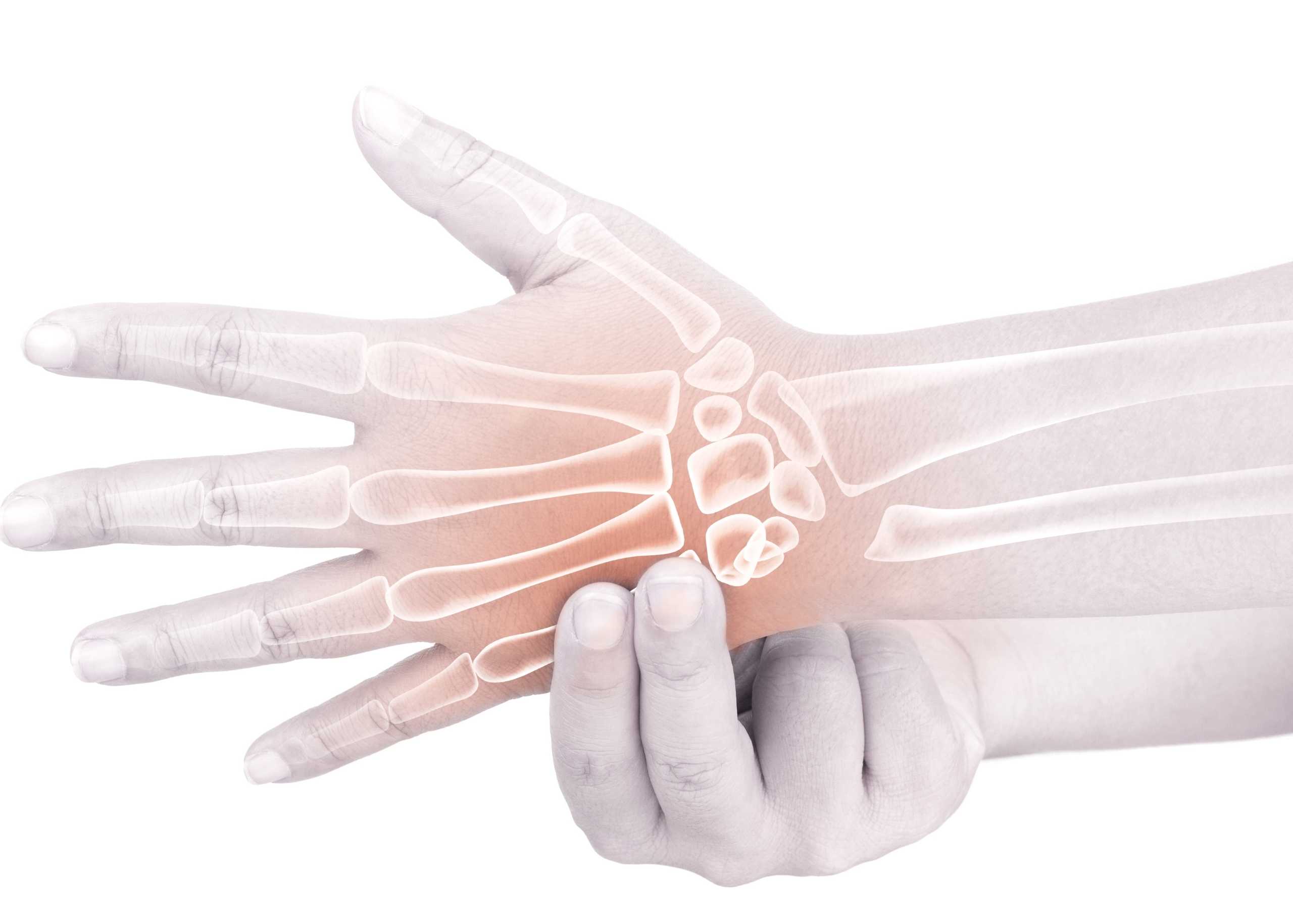
The symptoms of thumb arthritis include pain, swelling, and tenderness in the base of your thumb (the attachment area where it meets your wrist). You may also notice that your thumb becomes stiffer than normal when you try to move it. 

What Causes Thumb Arthritis? 

Thumb arthritis is caused by a breakdown of the joint cartilage and surrounding ligaments, which leads to excessive wear and tear on the joint. This causes pain and limited motion in the joint. 

The most common cause of thumb arthritis is osteoarthritis (OA) or degenerative joint disease (DJD). With OA, the protective cartilage layer that cushions your joints deteriorates over time due to age, injury, or overuse. This can result in bone rubbing against bone and causing pain.  

What are the Signs and Symptoms of Thumb Arthritis? 

The signs and symptoms of thumb arthritis include: 

  • Pain in your thumb joint when you move it or press on it 
  • Swelling in your thumb joint 
  • Stiffness in your thumb joint 
  • Decreased range of motion 
  • The enlarged or bony appearance of the joint at the base of your thumb 

How is Thumb Arthritis Diagnosed? 

Thumb arthritis is diagnosed through a physical exam, X-rays, and imaging tests. 

To diagnose thumb arthritis, a doctor will first perform a physical exam. They’ll look at the range of motion in your thumb and test the strength of your grip by asking you to pick up small objects with your thumb. This will help them determine if there are any changes in how your thumb moves or feels compared to before you were experiencing symptoms. 

Next, they’ll take an X-ray of your hand and wrist to assess whether changes in the bones or joints could be causing the pain. Suppose there are no obvious changes on the X-ray. In that case, they may recommend getting an MRI (magnetic resonance imaging) or CT (computed tomography) scan of your hand and wrist to provide more details about what might be causing your pain. 

How is it Treated? 

Thumb arthritis is treated with medication, physical therapy, and splinting. Based on the severity of the condition, surgery might also be required.  

Medications for thumb arthritis include corticosteroids, nonsteroidal anti-inflammatory drugs (NSAIDs), pain medications, and sometimes even steroids. These are used to reduce inflammation and pain. 

Physical therapy can help with range of motion exercises and strengthening exercises to keep your thumbs moving smoothly. 

A splint is better handmade that keeps your thumb in proper alignment while it heals. 

Several types of surgery can be performed on the thumb, depending on the nature of the injury. 

Arthrodesis: This procedure involves removing as much bone as possible from a joint before setting it into its new position, ensuring it maintains its alignment even after healing. Arthrodesis can treat arthritis and other conditions affecting your thumb joint. This old procedure is still useful for heavy workers. 

Trapeziectomy is another common type of surgery that involves removing only a portion of the bone rather than all of it. This type of procedure can be used to relieve pressure on nerves or tendons. 

Osteotomy involves breaking the bone (usually at the joint) and then realigning it to improve alignment and reduce pain. In some cases, the joint is fused after surgery.  

Arthroplasty: In this innovative surgery, the bone is replaced by an artificial implant that helps support your thumb during movement and prevents further damage from occurring because of osteoarthritis or other conditions affecting your joints. It preserves the length of the thumb and its strength. 

How Can I Prevent it from Getting Worse? 

To prevent the condition from worsening, you should start by taking care of your thumb joint. This means avoiding activities that may cause further damage to your joints. If you have a job that involves repetitive motion of your hands, try to reduce the amount of time spent performing those motions. You may also want to try using a splint at night to stabilize the joint while you sleep. 

You can also help prevent thumb arthritis from worsening by adopting an exercise routine that strengthens your hand and wrist muscles. Exercises help increase blood flow to your fingers and joints and improve flexibility, which will help reduce stress on the injured joint. 

Finally, suppose symptoms persist after these steps have been taken. In that case, it’s important to see a doctor specializing in hand conditions (such as a physical therapist or orthopedic surgeon) for further evaluation and treatment options. 

If you’re suffering from arthritis of your thumb, our hand surgeons at Burjeel SOS Hand Center in Dubai will work closely with you to determine which procedure suits your needs. We offer minimally invasive surgical procedures that allow us to treat patients without opening up their wounds or making long incisions during surgery. This means less pain, less scarring, and better results! 

Expert Hand Surgeon in Dubai

Dr. Marouane BouloudhnineConsultant Orthopedics Surgeon 


Years of Experience: 22

Nationality: France

Languages Known: Arabic, English, French, Italian 

Book Appointment

Press & Media

UAE becomes the first Gulf country to offer artificial joint replacement of the thumb

Our Experts

See More

Mammogram – What You Need To Know

What Is a Mammogram?

A mammogram is a radiographic examination of the breast that can help detect breast cancer in its early stages. It is recommended for women at average risk of developing breast cancer. 

When Should I Get a Mammogram?

The American Cancer Society recommends mammograms yearly for women aged 45 to 54. Women 55 and older should switch to mammograms every two years or can continue yearly screening.

How Do I Prepare for a Mammogram?

Burjeel Hospitals are outfitted with cutting-edge diagnostic equipment, such as the HOLOGIC 3DimensionsTM mammography system. You can visit one of the closest facilities to have breast cancer screening.

Bring a list of the locations and dates of past mammograms, biopsies, or other breast procedures, as well as your previous medical records, if you visit one of our facilities for the first time.

Schedule your mammogram around a time when your breasts are unlikely to be sore or swollen to minimize discomfort and obtain high-quality images. Avoid the week leading up to your period.

Do not apply deodorant, antiperspirant, powders, lotions, creams, or perfumes beneath your arms or on or under your breasts on the exam day. Some of these contain substances that can appear as white spots on x-rays.

Before a mammogram, discuss any recent changes or issues with your breasts with the doctor. (If you have symptoms, diagnostic mammography may be necessary to obtain images of the area of concern.)

Make sure the doctor is aware of any aspect of your medical history that could increase your breast cancer risks, such as surgery, hormone use, a family history of breast cancer, or a previous diagnosis.

What Will Happen During a Mammogram?

The breast tissue is stretched out during mammography by compressing the breasts between two firm surfaces. Then, an X-ray is used to acquire 3D images, which are shown on a computer screen and analyzed for cancer indicators.

Will a Mammogram Hurt?

A mammogram is a painless procedure. You won’t feel anything during your mammogram. The only thing you might notice is the pressure of the breast compression. Women experience discomfort during a mammogram due to pressure on the breast tissue, which can be uncomfortable for some women. The risk of pain depends on the sensitivity of your breasts. Most women do not notice discomfort during their annual mammogram unless they have sensitive breasts or have previously undergone breast surgery. If you are concerned about pain during your mammogram, let your doctor know before your appointment so they can apply additional gel to reduce friction between the breast and the film plate or compression paddle that is used during the procedure.

What Happens After the Test?

After the mammogram screening test, you’ll be asked to wait for your results. You will receive them either immediately or within a week. Still, it’s important to know that they can take up to two weeks if the radiologist needs more information to make a diagnosis.

Suppose there are concerns about your mammogram result. In that case, you may be asked to come back for another appointment or for further tests such as ultrasound or biopsy (which involves taking cells from the tumor and examining them under a microscope).

Where Can I Get a Mammogram?

Our facilities are equipped with the latest mammography systems. The HOLOGIC 3DimensionsTM mammography system provides the highest image quality for breast imaging. It can deliver high-resolution images with greater detail than other systems, as well as provide more accurate detection and diagnosis of breast cancer.

A HOLOGIC 3Dimensions™ mammography system is a revolutionary technology that uses special software to create three-dimensional images of your breast tissue. The 3D images allow radiologists to visualize breast tissue more accurately than traditional film-based mammography, leading to earlier detection of abnormalities.

The HOLOGIC 3Dimensions™ system also features IQM™ software that provides real-time analysis and enhancement of all breast images taken during your exam, ensuring that only the clearest, most diagnostic images are reviewed by your radiologist.

Early Detection of Breast Cancer Can Improve Outcomes

Early detection of breast cancer can improve outcomes by helping to prevent the disease from spreading throughout the body. There are many options for early detection, including screening mammograms, breast self-exams, and clinical breast exams.

If you notice any changes in your breasts, such as lumps or swelling, you should make an appointment with your doctor as soon as possible.

If You are a Woman Above 40, it is Time to Get Your Annual Breast Cancer Screening.


    Breast Cancer – What You Need To Know

    What is Breast Cancer?

    Breast cancer is the most common form of cancer in women. It occurs when cells in the breast become abnormal and start to grow out of control. The abnormal cells can be either benign (not cancer) or malignant (cancer). Breast cancer can be found in one breast or spread from one part of the body to another.

    What are the Types of Breast Cancer?

    There are several types of breast cancer, including:

    Angiosarcoma: A type of sarcoma (cancer of the soft tissue) that develops in the lining of blood vessels.

    Ductal Carcinoma In-Situ (DCIS): A noninvasive breast cancer that begins in the milk ducts and has not spread to other body parts.

    Inflammatory Breast Cancer: A rare form of breast cancer that develops quickly and grows more rapidly than other types.

    Invasive Lobular Carcinoma: A breast cancer that starts in the milk-producing glands’ lobules (tiny sacs).

    Lobular Carcinoma In-Situ (LCIS): A condition where abnormal cells appear in the lobules but do not grow into tumors or spread to other parts of the body.

    Male Breast Cancer: A rare form of cancer that affects men with breasts, most often those with Klinefelter syndrome or testicular feminization syndrome.

    Paget’s Disease of the Breast is a rare form of breast cancer in which the breast tissue becomes inflamed or infected, leading to a buildup of calcium deposits. It can also lead to changes in the skin, including thickening and discoloration.

    Recurrent Breast Cancer. Cancer cells return after treatment and surgery to remove them, causing more lumps or pain in the breasts.

    What are the Symptoms of Breast Cancer?

    It’s normal to be concerned about the symptoms of breast cancer. The most common symptom is a lump or mass in the breast, but there are others. Some of the other symptoms include:

    • lumps in lymph nodes (glands)
    • nipple discharge that is not milk
    • dimpling or puckering of the skin around the nipple
    • redness or flaking of the nipple
    • pain in any area of the breast, including the nipple
    • rash on your chest or under your arm
    • change in size or shape of your breast

    What are the Stages of Breast Cancer?

    Breast cancer is categorized into four stages based on the extent of the disease and the likely outcome.

    Stage I: The tumor is less than 2 centimeters across and has not spread to lymph nodes.

    Stage II: The tumor is between 2 and 5 centimeters across and has not spread to lymph nodes.

    Stage III: The tumor is greater than 5 centimeters across or has spread to one or more lymph nodes but not to distant organs.

    Stage IV: The tumor has spread to other body parts beyond the breast.

    What are the Risk Factors for Breast Cancer?

    Some of the risk factors for breast cancer include:

    Age—it’s most common in women over age 50, but it can occur at any age.

    Family History of Breast Cancer—if your mother or sister has had breast cancer, you are at higher risk of developing the disease yourself.

    Genetics—Women with a first-degree relative (mother, sister, daughter) who have had breast cancer are at greater risk of developing breast cancer. Women with BRCA1 or BRCA2 gene mutations are at higher risk of developing breast and ovarian cancers.

    Diet—certain foods may increase your risk of developing breast cancer; these include processed meats and red meats.

    Obesity—obese women are at higher risk for developing breast cancer than normal-weight women.

    How is it Diagnosed?

    It is often found through routine screenings such as mammograms and X-rays taken of your breasts. Mammograms can see early signs of breast cancer before you may feel any symptoms. 

    The American Cancer Society recommends that women get a mammogram every year starting at age 40 or in their 20s for those at high risk for developing this type of cancer A doctor will also recommend regular self-exams if you’re over 30 years old.

    It’s important to know what to expect during your diagnosis and treatment to feel prepared and supported throughout the process.

    Breast Exams

    A breast exam is an essential part of your breast health routine. You should schedule a visit with your doctor immediately if you notice any changes in your breasts, such as swelling or lumpiness. 

    Mammograms

    A mammogram is an X-ray of your breasts taken from different angles to check for abnormalities. A mammogram may lead doctors to perform additional testing, such as biopsies or ultrasounds, if they find something unusual during the procedure. These tests can help determine whether an abnormality is cancerous or benign (non-cancerous).

    Biopsy

    A biopsy removes tissue samples to examine them under a microscope for signs of cancerous cells.

    Breast Ultrasound

    An ultrasound uses sound waves to create an image of the breast tissue. Ultrasounds are usually recommended for women with dense breasts or who are pregnant because these conditions can make mammograms difficult to read accurately. 

    Breast MRI

    An MRI scan can also evaluate a woman’s breasts for signs of cancer or other conditions, such as a lump or tumor. MRI machines use strong magnetic fields and radio waves to create detailed images of soft tissues such as your breasts.

    How is it Treated?

    There are several treatment options that your doctor may recommend. These include surgery, radiation therapy, chemotherapy, hormone therapy, and targeted therapy.

    Surgery is the most common form of treatment. This procedure removes all or part of the tumor and any surrounding tissue. Your doctor may also take out lymph nodes if they think it is necessary.

    Radiation therapy uses high-energy beams to kill cancer cells to prevent them from growing back. It can be done alone or with other chemotherapy or hormone therapies.

    Chemotherapy is another treatment that uses drugs to kill cancer cells in the body and slow down their growth so they don’t spread as quickly. This type of treatment is often used with surgery or radiation therapy along with other treatments such as a hormone or targeted therapies, depending on the breast cancer you have been diagnosed with.

    Targeted therapies help destroy specific types of cells within your body by targeting certain proteins that are present on those cells only; this allows them to be destroyed without harming other healthy tissues nearby, which would happen if using traditional chemotherapy methods instead (such as most conventional chemo drugs).

    At Burjeel, we believe that cancer care is about treating the whole person, including their physical and emotional well-being. That’s why we offer a comprehensive range of services from leading oncology and palliative care experts.

    We are an ESMO Designated Centre of Oncology and Palliative Care. Our multi-disciplinary team of American and UK board-certified doctors includes medical oncologists, radiation oncologists, surgical oncologists, plastic surgeons, and palliative care specialists who work together to provide patients with the best possible care based on their needs.

    Early Detection Saves Lives. Get Screened Today!


      Gastroesophageal Reflux Disease (GERD) – Symptoms, Causes & Treatment

      Gastroesophageal Reflux Disease (GERD)

      Gastroesophageal reflux disease (GERD) is a digestive disorder in which acidic stomach fluids or food and fluids back into the stomach’s esophagus. GERD affects people of all ages, from infants to senior citizens. Asthmatics are at a higher risk of developing GERD.

      Causes of Gastroesophageal Reflux Disease

      Acid reflux, or the reflux of non-acid stomach material, frequently occurs and leads to GERD. A muscular band called the lower esophageal sphincter relaxes as you swallow, allowing food and drink to pass into your stomach. The sphincter then once again closes.

      Symptoms of Gastroesophageal Reflux Disease

      The symptoms of GERD include, 

      • Heartburn 
      • Regurgitation 
      • The feeling of food caught in your throat 
      • Coughing 
      • Chest pain 
      • Vomiting 
      • Sore throat and hoarseness   

      Prevention of Gastroesophageal Reflux Disease

      The occurrence of GERD can be minimized or prevented by,

      • Maintaining a healthy weight 
      • Quitting smoking 
      • Elevating the head of your bed 
      • By not lying down after a meal 
      • Eating food slowly and chewing thoroughly 
      • Avoiding foods and drinks that trigger reflux (e.g., soda, tomato juice, etc.)
      • Avoiding tight-fitting clothing    

      Diagnosis of GERD

      Based on your medical history, signs, and symptoms, as well as a physical examination to rule out other conditions or confirm the diagnosis of GERD, your doctor may be able to determine whether you have GERD, 

      • Upper endoscopy 
      • Ambulatory acid PH probe test
      • X-ray of the upper digestive system 
      • Esophageal manometry 

      Risk Factors of GERD

      The risk factors of GERD include, 

      • Exposure to cigarette smokes 
      • Hiatal hernia 
      • Medication and supplements 
      • Previous bariatric surgery (eg-metabolic surgery for weight loss)

      If you are suffering from GERD, it is essential to seek treatment as soon as possible. At Burjeel Hospital, MHPC, we offer comprehensive care for GERD. We can help you manage your condition with the help of our gastroenterology experts, who will work with you to develop an individualized plan to reduce symptoms while preserving your quality of life.

      Exercises For Osteoporosis

      Exercises for osteoporosis are the best form of prevention and treatment. Many exercises can be performed by anyone, no matter what age or level of fitness they are at.

      What is Osteoporosis?

      Osteoporosis is a condition where a person’s bones lose calcium and other minerals, which makes them fragile (breaking). According to a research study, osteoporosis and fragility fractures are an expensive human and socioeconomic burden everywhere in the world.

      Is it Safe to do Exercises if you Have Osteoporosis?

      Even with osteoporosis, it is never too late to begin a bone-healthy exercise program. You may be concerned that being active increases your chances of falling and breaking a bone. However, the opposite is true. 

      A regular, well-designed exercise program may aid in the prevention of falls and fractures. This is because exercise strengthens bones and muscles while improving balance, coordination, and flexibility. This is critical for people who have osteoporosis.

      What Are the Advantages of Exercise for People Who Have Osteoporosis?

      Fractures are more likely to occur when sedentism, poor posture, balance, and weak muscles are present. Numerous approaches, including exercise, can help persons with osteoporosis improve their health. Other benefits include, 

      • Reduction in bone loss
      • Increased physical fitness since the residual bone tissue is preserved.
      • Strengthened muscles
      • Enhanced reaction time and increased mobility
      • Improved coordination and balance
      • Decreased chance of bone fractures brought on by falls
      • Pain relief, increased vigor, and better mood

      What Kind of Exercises Should I Do?

      Before starting a new exercise regimen, speak with your doctor and physical therapist. They can advise you on what is safe for your level of exercise, overall health, and stage of osteoporosis.

      There isn’t a single fitness program that works for everyone with osteoporosis. Your routine should depend on your age, the severity of your osteoporosis, and the medication you are taking right now.

      Additional medical diseases such as arthritis, cardiovascular or pulmonary illness, and neurological issues affect your ability and fitness. 

      Improving bone density or preventing falls should be the major objectives of your exercise regimen. Mix specific balance exercises with weight-bearing aerobic and muscle-building (resistance) training is preferable.

      What Exercises are Recommended for Osteoporosis?

      • Exercising with weights. You do them on your feet, which forces your bones and muscles to work against gravity to keep you upright. Your bones respond to weight by strengthening and building themselves. Weight-bearing exercise is classified into two types: high-impact and low-impact.
      • Resistance training with dumbbells and barbells, elastic band resistance, body-weight resistance, or weight-training machines
      • Tai chi and other posture, balance, and body strength exercises

      What Exercises/Activities Should People with Osteoporosis Avoid?

      A person who has osteoporosis has weakened bones that are prone to breaking. They should avoid engaging in activities such as: 

      • Forward spine flexion with weighted loads, such as abdominal sit-ups. Increases the likelihood of you falling. It needs quick, strong action unless taught gradually as part of a progressive program. 
      • A powerful twisting motion, like a golf swing, is necessary unless the person is accustomed to such movements.

      Are There Any Recommendations for the Frequency and Duration of Exercise?

      The precise amount of exercise required for people with osteoporosis is unknown. However, guidelines recommend:

      • 45 to 1 hour of aerobic exercise twice or three times a week
      • Each resistance training session should include exercises to develop the lower limb, trunk, and arm muscles. Resistance training should be done twice or three times a week. Eight to ten times should be given to each exercise.
      • Exercises to improve your balance should be done twice a week for a few minutes at a difficult level. For safety reasons, always ensure you have something you can grip on if you overbalance something.
      • Exercises that promote flexibility include stretching.

      You must consistently perform your activities throughout time to lower your risk of suffering a bone fracture.

      What Else Can You Do to Improve Bone Health?

      Exercise is an essential component of any osteoporosis treatment plan. 

      Before beginning a new exercise program, consult with your doctor. Expert advice can be obtained from physiotherapists and other exerc ise professionals.

      Always begin your exercise program at a low level and gradually progress. 

      Exercising too quickly and vigorously may increase your risk of injury, including fractures. 

      Also, talk to your rheumatologist or dietitian about ways to boost your calcium, vitamin D, and other essential nutrient intakes. They may suggest that you take supplements. Smoking and excessive alcohol consumption are both terrible for your bones.

      Exercising for Osteoporosis is a significant part of managing your condition, and you should start working on it as soon as possible. If you have not yet begun an exercise routine, consult one of our expert Physiotherapists at Burjeel Hospital, Abu Dhabi, who can help you get started.

      Alzheimer’s Disease – Symptoms, Causes & Treatment

      Alzheimer’s disease is a brain disease that gradually deteriorates thinking and memory abilities and the ability to perform even the most basic tasks. The majority of Alzheimer’s patients have their initial symptoms later in life. According to experts, around 55 million people in the world who are 65 or older have dementia, according to various estimates. 

      The most prevalent type of dementia, Alzheimer’s, maybe a factor in 60–70% of cases. In today’s world, it is one of the primary causes of disability and dependency among older people and the seventh most significant cause of death among all diseases.

      For those living with dementia, their caregivers, families, and society, dementia has physical, psychological, social, and economic effects.

      How is the Brain Affected by Alzheimer’s Disease?

      Scientists fully understand the intricate brain alterations associated with Alzheimer’s disease. Before symptoms show, changes in the brain may start ten years or earlier. The brain experiences harmful alterations at this early stage of Alzheimer’s, including aberrant protein buildups that result in amyloid plaques and tau tangles. 

      Formerly healthy neurons stop working, lose connections to neighboring neurons, and eventually die. Numerous other intricate brain alterations also influence Alzheimer’s disease.

      The hippocampus and the entorhinal cortex, two brain regions crucial for memory formation, appear to be the first areas of the brain to sustain injury. Additional brain regions are damaged and start to shrink as more neurons die. When Alzheimer’s reaches its most advanced stage, the damage is extensive, and the brain tissue has greatly diminished.

      What are the Warning Signs and Symptoms of Alzheimer’s?

      One of the initial indications of Alzheimer’s-related cognitive decline is frequent memory issues. Some individuals with memory issues experience minor cognitive impairment (MCI). 

      People with MCI experience more memory issues than is typical for their age. Nevertheless, their symptoms do not impact how they conduct their daily lives. MCI has also been associated with problems with movement and the sense of smell. Alzheimer’s disease is more likely to affect older MCI patients, but not all of them do. Some people might even resume their regular thinking. Individuals with Alzheimer’s experience different initial symptoms. 

      Many people believe that the very early stages of the disease may be indicated by a deterioration in non-memory components of cognition, such as word finding, vision/spatial problems, and impaired thinking or judgment. 

      To identify early abnormalities in the brains of persons with MCI and cognitively healthy individuals, researchers are examining biomarkers, which are biochemical indications of disease discovered in blood, cerebrospinal fluid, and brain imaging. They might be more susceptible to Alzheimer’s. Before these methods can be widely and regularly used to diagnose Alzheimer’s in a physician’s office, more study is required.

      What are the Different Stages of Alzheimer’s Disease?

      Mild Alzheimer’s Disease

      Memory loss and other cognitive problems get worse as Alzheimer’s gets worse. Wandering and getting lost can be a problem, as can having difficulties managing money and paying bills, asking questions repeatedly, taking longer to do everyday duties, and changing their personality and conduct. In this period, patients are frequently diagnosed.

      Moderate Alzheimer’s Disease

      This stage of the disease involves brain damage to the regions responsible for language, cognition, conscious thought, and sensory processing, including the capacity to recognize sounds and scents. 

      As disorientation and memory loss worsens, it gets harder for people to identify their loved ones. They might be unable to adapt to new circumstances, learn new things, or perform complex chores like getting dressed. At this stage, individuals may act impulsively and have hallucinations, delusions, and paranoia.

      Severe Alzheimer’s Disease

      Plaques and tangles eventually cover the entire brain, and the brain’s tissue begins to diminish dramatically. People with advanced Alzheimer’s cannot communicate and rely on others to care for them. As the body slows down at the end of life, the person may spend most of the time in bed.

      What Causes Alzheimer’s Disease?

      Scientists have made enormous strides toward understanding Alzheimer’s disease in recent years, and the impetus is still building. Even still, researchers are unsure of the exact causation of Alzheimer’s in most cases. A genetic mutation might be the root cause of early-onset Alzheimer’s in affected individuals.

      A complicated succession of brain alterations may occur over many years, leading to late-onset Alzheimer’s disease. Genetic, environmental, and lifestyle factors are likely the root causes. Depending on the individual, each of these characteristics may play a different role in elevating or lowering the chance of developing Alzheimer’s.

      How is Alzheimer’s Diagnosed?

      Doctors employ various techniques and instruments to assess whether a patient with memory issues has Alzheimer’s. For an Alzheimer’s diagnosis, clinicians may:

      • Inquire about the person’s general health, prescription and over-the-counter medications, food, previous medical issues, capacity to carry out daily tasks, and any changes in behavior or personality, together with a family member or friend.
      • Tests for language, counting, problem-solving, and memory should be conducted.
      • Perform standard medical examinations, such as blood and urine testing, to look for further potential reasons for the issue.
      • To confirm an Alzheimer’s diagnosis or rule out other potential causes of symptoms, conduct brain scans using computed tomography (CT), magnetic resonance imaging (MRI), or positron emission tomography (PET).
      • These tests may be repeated to inform clinicians about how the patient’s memory and other cognitive abilities evolve.

      People who experience difficulties with memory and thinking should consult their neurology doctor to determine whether the symptoms are caused by Alzheimer’s disease or something else, such as a stroke, tumor, Parkinson’s disease, sleep disturbances, medication side effects, an infection, or another form of dementia. Some of these ailments might be treatable and even curable.

      If Alzheimer’s disease has been identified, starting treatment as soon as feasible in the disease course may assist in maintaining everyday functioning for a while. A timely diagnosis aids families in making plans. 

      They can deal with monetary and legal issues and potential safety concerns, learn about living situations, and create support networks.

      How is Alzheimer’s Treated?

      Due to the complexity of the disease, it is doubtful that any medication or other treatment would completely cure everyone with Alzheimer’s. 

      Alzheimer’s disease is currently being treated with an emphasis on managing behavioral symptoms, treating the underlying disease process, and assisting patients in maintaining mental function. 

      What are the Medications to Maintain Mental Function in Alzheimer’s Disease?

      The US Food and Drug Administration (FDA) has approved several drugs to treat Alzheimer’s symptoms. The signs of mild to moderate Alzheimer’s disease are managed using donepezil, rivastigmine, and galantamine. 

      The rivastigmine patch, memantine, donepezil, and medicine that combines the two are all used to treat mild to severe Alzheimer’s symptoms. All these medications function by controlling neurotransmitters, substances that communicate between neurons. 

      They might aid in treating some behavioral issues and symptom reduction. However, the underlying illness process is unaffected by these medications. They may be helpful for a brief period and are successful for some people but not everyone.

      What are the Medications to Treat the Underlying Alzheimer’s Disease Process?

      The FDA has approved aducanumab as the first Alzheimer’s disease disease-modifying medication. The medication helps to reduce the brain’s amyloid buildup. It might slow the disease’s course. It has not yet been demonstrated that it influences clinical outcomes, such as the development of dementia or cognitive decline.

      A physician or neurologist would probably run tests, such as a PET scan or a cerebrospinal fluid analysis, to check for signs of amyloid plaques and determine if the patient is a good candidate for the proposed course of treatment.

      How to Manage the Alzheimer’s Disease Behaviour?

      Sleeplessness, wandering, agitation, anxiety, and hostility are typical behavioural signs of Alzheimer’s disease. Scientists are discovering the causes of these symptoms and researching new drug and non-drug management strategies.

      According to research, addressing behavioural symptoms can help caregivers, and patients with Alzheimer’s disease feel more comfortable. Family and caregiver support for those with Alzheimer’s illness

      Alzheimer’s care can be physically, emotionally, and financially expensive. 

      It can be challenging to balance daily care responsibilities, family dynamics shifts, and choices regarding placement in a care facility.

      One crucial long-term approach is to educate yourself on the illness. 

      Programs that instruct families on how to handle complex behaviours and other caregiving issues, as well as the many stages of Alzheimer’s, might be helpful.

      The expert Neurologists at Burjeel Hospital provide strategies to help carers manage the stress of caring for a loved one with Alzheimer’s, including effective coping mechanisms, solid support systems, and respite care.

      For instance, maintaining an active lifestyle has both psychological and physical advantages. For some carers, finding a support group to join is a crucial lifeline. These support groups give caregivers a place to obtain emotional support, voice worries, share experiences, and acquire ideas.


      Our Expert Neurologist


      Dr. Halprashanth D.S

      Consultant Neurology

      Years of Experience : 11

      Nationality : India

      Languages Known : English, Urdu, Hindi, Kannada, Tamil

      Burjeel Hospital, Abu Dhabi

      Hand Fracture – Signs, Symptoms, and Treatment

      What is a Hand Fracture?

      A hand fracture is a break in one or more of the bones that make up your hand. Hand fractures are most commonly caused by direct trauma to the hand, such as when you fall on your outstretched hand or catch yourself with your hands after tripping.

      Hand fractures can be either open (where the bone breaks through the skin) or closed (where the bone does not break through the skin). Hand fractures are typically treated with a cast to immobilize and protect the injured area while it heals.

      What are the Most Common Types of Hand Fractures?

      The most common types of hand fractures are:

      Colles’ Fracture: This is a break in your forearm’s lower end of the radius bone. It can happen when you fall on an outstretched hand or have an elbow injury and try to catch yourself by putting out your hand.

      Scaphoid Fracture: This is a break in the scaphoid bone located just above your wrist. It usually happens from falling on an outstretched arm or twisting your wrist too far while lifting something heavy.

      Monteggia Fracture: refers to dislocation of the radial head (proximal radioulnar joint) with fracture of the ulna.

      What are the Symptoms of a Hand Fracture?

      The symptoms of a hand fracture can vary depending on the severity of the injury. In general, if you experience any of the following signs and believe you may have suffered a fracture, contact your physician immediately:

      • Pain and swelling in the hand
      • Difficulty moving or using the hand
      • Deformity of the hand and/or fingers

      How is a Hand Fracture Diagnosed?

      A doctor diagnoses a hand fracture through a physical examination and X-rays. The doctor will examine your hand for tenderness, swelling, deformity, and bruising. They may also check for neurological damage or abnormal sensation in the fingers. In some cases, an MRI scan might be needed to determine the extent of the damage.

      How is a Hand Fracture Treated?

      A hand fracture is treated differently depending on the type of fracture.

      A simple fracture, which involves only one bone, is treated by placing a splint on the hand and wrist. The splint keeps the bones in place while they heal.

      Complex fractures are more severe and involve multiple bones or joints. They may require surgery to place pins and screws to hold the bones in place while they heal.

      In some cases, fractures can be treated with surgery alone. In these cases, it is essential to consult an expert hand surgeon about whether surgery is recommended for your specific condition.

      Hand fractures are common injuries for athletes and active people. They can be caused by a fall, an impact from a hard object or another person, or an accident that involves sudden braking. If you suspect you have a hand fracture and cannot move the injured finger or knuckle, or if your finger or knuckle is deformed, swollen, or discolored, you should seek medical attention immediately.

      Expert Hand Surgeons at Burjeel Hospital Dubai provide the best treatment for hand fractures. We will work with you to determine the best way to treat your fracture, whether that means surgical intervention or non-surgical treatment.

      Our Expert Hand Surgeons in Dubai

      Dr. Marouane Bouloudhnine

      Consultant Orthopedics Surgeon

      Years of Experience: 22

      Nationality: France

      Languages Known: Arabic, English, French, Italian

      Dr. Khalid Alawadi

      Consultant Hand & Plastic Surgeon (Specialized in Plastic, Reconstructive & Hand Surgery) (Visiting Doctor)

      Years of Experience: 21

      Nationality: UAE

      Languages Known: Arabic, English, German

      Dr. Jamil Al Jamali

      Consultant Hand Surgeon (Specialized in Plastic & Reconstructive Microsurgery) (Visiting Doctor)

      Years of Experience: 22

      Nationality: Germany

      Languages Known: Arabic ,English, German

      Our Experts

      See More

      Tooth Discoloration – Symptoms, Causes & Treatment

      Have you looked in the mirror while getting ready for a big day and noticed that your teeth are not as white as you would like them to be? While you might know how to “fix” it, let’s talk about what causes your teeth to be stained and how to prevent it.

      What Causes Tooth Discoloration?

      Aging. As you age, the transparent enamel surface of your teeth may wear down, disclosing the yellowish color of the underlying dentin. 

      Poor Oral Hygiene. This causes the accumulation of food debris and plaque formation, a thick layer rich in bacteria. These factors can cause the teeth to look stained and yellow.

      Tooth Decay. Poor oral hygiene can lead to decay of teeth which starts as white patches and then turns brown or black. 

      Habits. Excessive intake of tea or coffee and smoking can cause brown staining of the teeth.

      Stress. Stress affects most body functions, and among that, it affects the normal flora of your mouth, which results in the activation of some kinds of bacteria, which will result in dotted black stains on the teeth at the gum line.

      Fluoride. Too much fluoride can cause white spots. This is called fluorosis, and it happens when teeth are exposed to much fluoride from drinking water or excess use of fluoridated mouthwashes or toothpaste.

      Improper Dental Treatments. Amalgam filling (silver fillings) can cause the tooth to look black. When root canal treatment is done, tooth color changes to yellow, brown, or even black in some cases.

      Antibiotics. Exposure to some antibiotics during tooth formation, like in the mother’s womb or at an early age, can cause yellow to green lines on the teeth. This kind of stain is internal and needs more invasive treatment to be covered.

      How to Prevent Teeth Staining?

      Prevention is key to healthy teeth. For that, try to minimize the intake of colored beverages, avoid smoking, reduce stress, maintain a good oral hygiene routine, and visit the dentist regularly.

      How is Tooth Discoloration Treated?

      There are multiple options to have a brighter smile and whiter teeth.

      Brushing your Teeth. when you miss brushing for a few days, your teeth will have a yellow plaque layer. Once you brush, you will see the difference.

      Scaling and Polishing in the clinic will remove all the calculus, plaque, and black stains, leaving your teeth looking whiter and brighter.

      In-Office Teeth Whitening. It’s a professional way of removing the stains absorbed by your teeth over the years, disclosing the white and bright teeth you have,

      Proper Dental Treatment. We might need to replace fillings, do internal bleaching or do root canal treatment again to remove the discolored improperly done root canal filling materials

      Crowns and Veneers are used in case of internal staining caused by the use of antibiotics and for the aged teeth to replace the lost tooth structure and get the white smile you deserve.

      Having a bright smile is a matter of having a good oral hygiene routine, and tooth discoloration can affect how you feel about your smile and oral health. Treatments and prevention methods exist to help you feel more confident. Our Expert Dentists at Burjeel MHPC provide various services to help you maintain the health of your teeth and gums.

      Our expert Dentists will work with you to determine the best approach for restoring your tooth’s natural color and shape and preventing future discoloration. From dental crowns to veneers, we have the skills necessary to provide quality treatment that is both safe and effective.

      Cardiac Rehabilitation Program

      What is Cardiac Rehabilitation? 

      Cardiac rehabilitation is a program to help patients with heart disease or surgery recover their strength and fitness. It can also help people with other heart conditions, such as diabetes, high blood pressure, or sleep apnea. Cardiac rehabilitation programs usually involve 

      • exercise training, 
      • education about healthy nutrition and behavior change, 
      • support from other patients in the program, and 
      • sometimes psychological counseling. 

      A cardiac rehabilitation program can be tailored to meet your needs and meet at times that are convenient for you. The most important part of cardiac rehab is exercise training. This helps your body get stronger and more efficient at managing its health. 

      Who Needs Cardiac Rehabilitation? 

      Cardiac rehabilitation program is not only for people who have had heart surgery but also for those who have had a heart attack or other conditions that affect their hearts. These include: 

      • Aneurysm (a bulge in the wall of an artery) 
      • Arrhythmia (irregular heartbeat) 
      • Heart attack 
      • Heart failure 
      • High blood pressure 

      What does Cardiac Rehab Involve? 

      Cardiac rehab starts with an initial evaluation by a doctor. The goal of cardiac rehabilitation is to help patients learn how to manage their heart disease and prevent future complications. Cardiac rehabilitation includes several different types of interventions, including: 

      Behavioral interventions focus on changing the patient’s behaviors to reduce their risk for future complications. Behavioral interventions include smoking cessation programs and weight loss programs. 

      Dietary interventions teach patients healthy eating habits to help them manage their heart disease symptoms and prevent future complications. 

      Exercise training focuses on helping patients learn how to exercise safely to improve their cardiovascular fitness levels and reduce the risk for future complications. 

      After the initial evaluation, you will meet with your cardiologist. They will discuss your medications, how much activity you should be doing, and how often to exercise. 

      You will also meet with an exercise physiologist who will design an exercise program that meets your needs and goals. The goal is to gradually increase the intensity of your physical activity over time while staying within safe limits based on your current health status. 

      Cardiac rehab sessions typically last six weeks but can vary depending on your condition and needs. Cardiac rehab usually involves two types of exercise: aerobic and resistance training.  

      Aerobic exercise includes walking on a treadmill, using an elliptical machine, or running on a track.  

      Resistance training involves lifting weights to build muscle mass. Cardiac rehab also includes education on healthy diet and lifestyle choices, such as quitting smoking and managing stress levels. 

      What are the Advantages of Cardiac Rehabilitation? 

      Some of the advantages of cardiac rehab include: 

      • Stress management: Cardiac rehab teaches you how to manage stress through exercises, which can help reduce your blood pressure and keep your heart healthy by reducing the amount of cortisol in your body. 
      • Improved fitness: Cardiac rehab improves your fitness level by getting you moving again and engaging in activities that increase endurance, strength, flexibility, and balance. This is especially important if you’ve recently had an illness or surgery because it’s crucial to stay active when recovering from these procedures so that you don’t lose too much muscle mass or become too weak. 
      • Improved diet: Cardiac rehab helps you develop healthy eating habits so that you’re not just eating whatever foods are convenient for when you’re hungry or tired—you’re ingesting nutrients from foods that will benefit your body long-term. 

      What Happens After Cardiac Rehab? 

      After cardiac rehab, you will continue to practice your new healthy habits. You will be advised to continue with your prescribed exercise program and any medication.  

      Your cardiologist and physiotherapist will continue to monitor your health and may recommend further treatment if necessary. 

      How to Stay Fit After a Heart Attack? 

      After a heart attack, staying fit is essential to improve your health and reduce the risk of a second heart attack. Getting back into your routine as soon as possible is crucial, including exercise. 

      Here are some tips for staying fit after a heart attack: 

      • Start with walking. Walking is the easiest way to move again, primarily if you’re not used to exercising regularly. It can also help you lose weight and lower blood pressure. Walking 30 minutes daily will help you regain shape faster than other exercises. 
      • Increase your activity gradually until you do 30 minutes of moderate exercise five times per week (for example) or 20 minutes of vigorous activity three times per week (like running). You may need to take it slow at first—even just getting out of bed in the morning and walking around may be enough for now! You should be able to add more time as long as it doesn’t cause pain or fatigue. 
      • Don’t forget about strength training! After a heart attack, your body will be weaker than before because it needs more oxygen due to damaged muscle tissue that needs repair; therefore, strength training is essential. 

      Cardiac rehabilitation can save your life after a heart event like a heart attack. It helps reduce the risk of dying from heart disease and the risk of having another heart event. It enables you to get back to being an active community member. 

      Researchers at Harvard Medical School reported that cardiac rehab reduces heart-related mortality by 20%, hospital readmissions by 28%, and increases the patient’s quality of life, ability to carry out daily activities, and overall mental well-being. 

      Our experienced physiotherapists are dedicated to helping you recover from a heart attack or surgery. They will guide you through a comprehensive program of exercises, which have been tailored specifically for your needs to help you achieve your goals—whether that’s walking again or simply feeling better. 

      We believe in empowering our patients and helping them regain control over their health. If you’re looking for an expert team of physiotherapists who can help support you on your journey back to wellness, look no further than Burjeel Hospital, Abu Dhabi. 

      Anal Fistula – Symptoms, Causes & Treatment

      What is Anal Fistula?

      Anal Fistula is a condition where the skin around the anus becomes inflamed and develops a tunnel-like opening. This tunnel can lead to a chronic infection that may require surgery to repair. This tunnel allows bacteria to enter the body and can cause severe pain. Anal fistulas are most common in people who have Crohn’s disease. Still, they can also occur in people with other conditions such as ulcerative colitis or diverticulitis.

      What Causes of Anal Fistula?

      Anal fistulas are usually caused by chronic inflammation and infection in the lining around the anus and rectum, which can result from various factors, including:

      • Constipation
      • Diarrhea
      • Infection of the lining around the anus (anal fissure)
      • Inflammation of the tissue inside the rectum (internal hemorrhoids)

      What are the Symptoms of an Anal Fistula?

      Symptoms of an anal fistula include:

      • Painless discharge from the anus that may be clear, cloudy, or bloody
      • Pain in the lower abdomen, bottom, or perineum
      • Swelling in the area around the anus
      • Persistent itching or burning sensation around the anus
      • Painful bowel movements

      How is Anal Fistula Diagnosed?

      Anal fistulas are usually diagnosed by a doctor and then confirmed with tests. The following tests may be used:

      Physical examination: Thedoctor will examine the anus and rectum to check for abnormalities. They may also ask about the symptoms and do a rectal examination to find out more about the exact location of the fistula.

      Rectal exam: A proctoscopy is where a special telescope with a light on the end is used to look inside the anus and rectum. It helps detect an anal fistula so treatment can start as soon as possible. Sometimes, the doctor may recommend additional tests as part of the evaluation. These tests include an ultrasound scan, MRI scan, or CT scan.

      Ultrasound scan: This test uses sound waves to create an image of internal structures within the body, such as organs or blood vessels. It may be used during surgery if there is no other way to determine if there’s an anal fistula present and may help guide doctors when trying to repair one surgically by showing them where it is located in relation to nearby organs such as nerves or blood vessels that could otherwise be damaged during surgery.

      What are the Risk Factors for Anal Fistula?

      The risk factors for anal fistulas include:

      • Age: The older an individual is, the higher their risk for developing new anal fistulas.
      • Gender: Women are more likely to develop anal fistulas than men.
      • Diabetes: This condition increases the risk of developing an anal fistula and other complications related to Crohn’s disease or ulcerative colitis.
      • Anal Fissure is a tear in the lining of the anus.
      • Anal Abscess is an infection in the area between the anus and the rectum.
      • Crohn’s Disease or Ulcerative Colitis, diseases that cause inflammation of the digestive tract.
      • Diarrhea can cause chronic constipation or hard stool that sticks and causes pain when it passes through the anus.
      • Hemorrhoids are swollen blood vessels around the anus that cause pain and bleeding.

      What are the Types of Anal Fistula?

      There are four main types of anal fistulas.

      • Intersphincteric Fistulas occur at the level of the internal sphincter, which is located between the internal and external sphincters.
      • Transphincteric Fistulas occur at the level of the external sphincter, which is located between the internal and external sphincters.
      • Suprasphincteric Fistulas occur above both internal and external sphincters, usually as a result of trauma to the anus or rectum.
      • Extrasphincteric Fistulas occur outside both internal and external sphincters, usually as a result of trauma to the anus or rectum.

      What are the Treatments Available for Anal Fistula?

      Anal fistulas are usually treated by a general surgeon, a colorectal surgeon, or a proctologist (an expert in diseases of the anus and rectum). Treatment for anal fistulas may include the following:

      Fistulotomy involves cutting out the internal portion of the fistula with a surgical knife or laser beam, allowing normal tissue to grow in its place. It’s usually performed under general anesthesia and requires an overnight stay in the hospital.

      Seton Techniques are used to treat anal fistulas. Setons are fine wires placed in the fistula tract to drain pus and fluids from the anal canal. The seton is removed after several months when healing has occurred.

      The Advancement Flap Procedure involves creating a new passage for fecal matter to exit the body by moving tissue from one area to another. The advancement flap procedure is typically performed in conjunction with other procedures, such as a posterior sphincterotomy or surgical removal of affected tissues. The advancement flap procedure may also be referred to as a mucosal advancement flap procedure.

      The Ligation of Intersphincteric Fistula Tract (LIFT) procedure is a minimally-invasive surgery that involves removing a portion of the intersphincteric fistula tract and then closing it with absorbable suture closure.

      Endoscopic Ablation is a minimally-invasive procedure that uses lasers to remove the damaged tissue from around the anus and close off the tunnel. This allows the area to heal and prevents further infection from occurring.

      Laser Surgery for anal fistula is a minimally invasive procedure that can be used to treat the condition. It can be performed under local or general anesthesia and often involves only one or two small incisions in the affected area. A laser beam is then used to seal off the openings of the fistula, which helps prevent infection from spreading and relieving symptoms.

      Fibrin Glue is a medical adhesive used to treat anal fistulas. Fibrin glue is made from a clotting protein called fibrinogen, extracted from human blood. Fibrin glue can be injected into the anal canal to seal off the fistula and stop the leaking of fecal matter, which causes the symptoms of this condition.

      FAQ’s

      How serious is a fistula?

      A fistula is a serious medical condition that can lead to surgery and other complications.

      How serious is a fistula?

      A fistula is a serious condition that can cause many problems for an individual’s health, both short-term and long-term. It’s important to understand what causes fistulas, how they’re treated and managed, and the potential complications.

      Can a fistula heal on its own?

      Fistulas can heal on their own, but the rate of healing varies depending on the severity of the condition. Fistulas are not always life-threatening but can be incredibly uncomfortable and require treatment. If an individual has a fistula, they should see their doctor find out how to treat it.

      Is fistula a major surgery?

      Fistula is not major surgery. The procedure is often performed under local anesthesia; most patients can go home the same day. However, some patients may need to stay in the hospital overnight.

      How long does fistula surgery last?

      Fistula surgery lasts anywhere from 30 minutes to 2 hours. The length of the procedure depends on how many surgeries an individual has, what type of fistula a persona has, and how complicated the case is.

      How painful is fistula surgery?

      Fistula surgery is a minimally invasive procedure. It is typically done under general anesthesia, and the patient will spend one to two hours in the recovery room after the surgery is complete.

      What food should be avoided in fistula?

      One way to avoid worsening a fistula is to avoid foods containing indigestible carbohydrates like roughage or fiber. These include wheat bran, whole wheat bread, cereals, beans and lentils, nuts, and seeds. The problem with these foods is that they can be difficult to digest, which can contribute to bacterial overgrowth in the small intestine, leading to inflammation in the area around the fistula or diverticular abscesses. Fistula pain is a burning sensation that can be felt in the lower abdomen, groin, or rectum. It may be experienced as a constant dull ache or as sharp pains that come and go.

      What does fistula pain feel like?

      Fistula pain can vary from person to person. Some patients may feel a burning sensation, while others may feel more of an ache or aching sensation. The pain can differ depending on where the fistula is located and how long it has been there.

      How do you heal a fistula?

      Fistulas can be very painful, but there are several ways to heal them. If there is an open wound, it’s essential to keep it clean and covered with a bandage. One should also avoid walking around in public, as this could expose it to bacteria that can worsen the fistula. If one has an internal fistula, their doctor may prescribe antibiotics or other medications to help treat the infection. One should also consider undergoing surgery if symptoms don’t improve after using antibiotics for several weeks or months. The type of surgery performed will depend on what type of fistula an individual has.

      Our expert Proctologists can help manage anal fistula with a combination of medications, procedures, and lifestyle changes. We’ll work with you to identify the best treatment for your needs.

      Our Expert Proctologist

      Dr. Ibrahim Gamal

      Specialist General Surgeon

      Years of Experience : 10

      Nationality : Eygpt

      Languages Known : Arabic , English