Kidney Stones

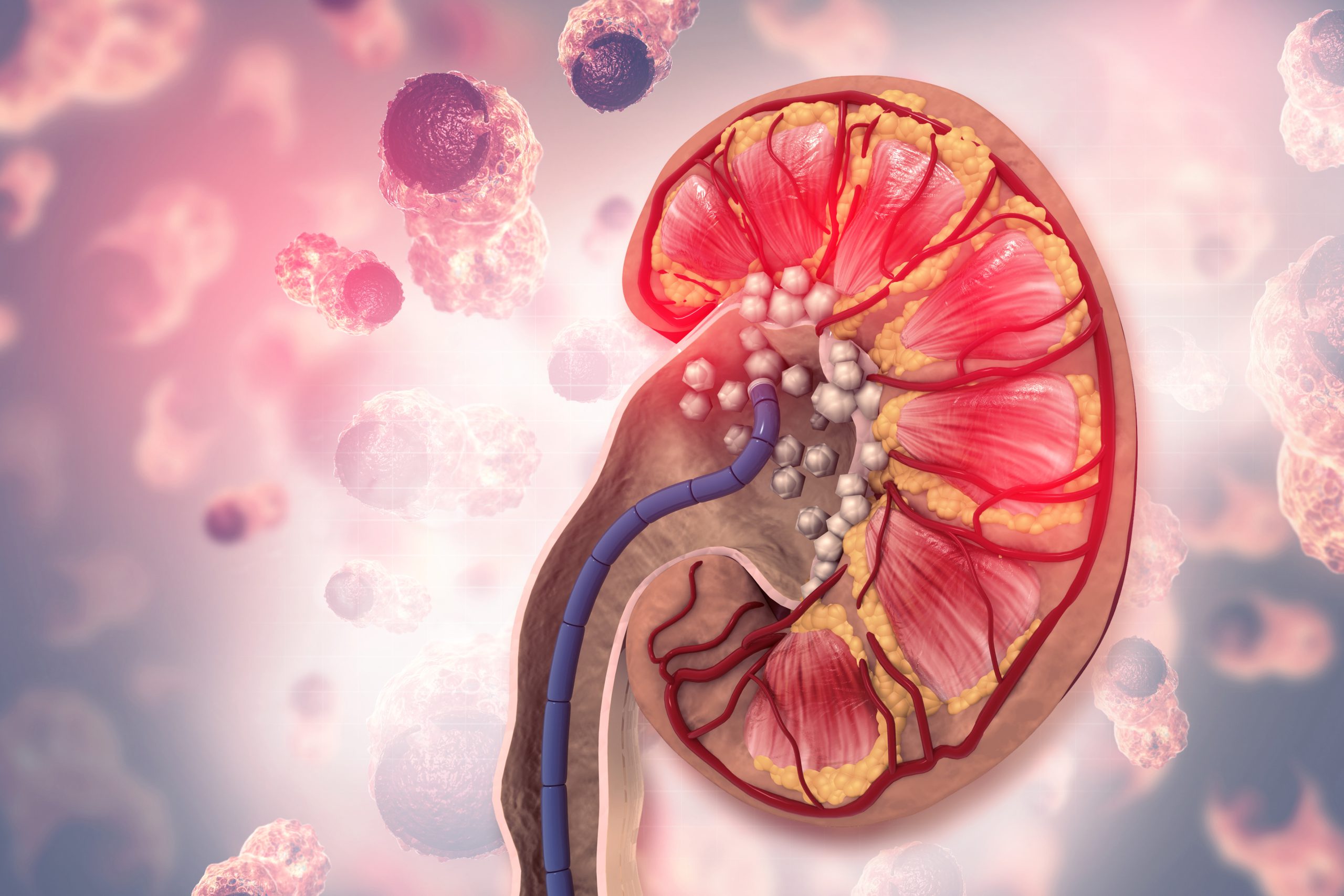
Kidney stones are hard deposits that form within the kidneys. They can cause a lot of pain and make it difficult to urinate. They are made from substances like calcium and can be as small as a grain of sand or as big as a golf ball. They often result from a buildup of minerals in the urine. The most common type of kidney stone is made up of calcium, but other substances may also be present. 

Most of them pass spontaneously within days or weeks, especially if you drink plenty of liquids and increase your water intake. However, if you have recurrent kidney stones, surgery might be necessary to remove them permanently or prevent their recurrence. It is essential to let your Urologist know if you’ve had any symptoms here. 

Types of Kidney Stones 

Kidney stones can be divided into four main types: calcium oxalate, calcium phosphate, uric acid, and struvite. 

  • Calcium oxalate stones are the most common type. They form when calcium combines with oxalate in urine
  • Calcium phosphate stones form when calcium combines with phosphorus to form crystals that then become hard material in your kidneys. Doctors don’t know why this happens 
  • Uric acid stones are caused by excess uric acid in your system. The acid makes its way into your urine and forms crystals there. These crystals bond together to create a stone that can block your urinary tract 
  • Struvite stones. A bacterial infection caused in your urinary tract or bladder 
  • Cystine: They are Rare. They occur in both men & women who have the genetic disorder cystinuria 

Causes of Kidney Stones

They occur when there is an imbalance in the number of minerals in the urine. The minerals crystallize and form crystals that stick together to form a stone. The crystals can become stuck in the kidney, ureter (the tube that connects the kidneys to the bladder), or bladder. This causes pain and other symptoms, such as nausea and vomiting, blood in urine, frequently urinating and feeling like you need to go. Still, nothing comes out when you do, needing to use the bathroom urgently but having trouble getting there quickly enough. 

Warning Signs of Kidney Stones 

The following are some of the warning signs: 

  • Pain in your side or back that is worse when you urinate, or pain while passing urine 
  • Bloody or cloudy urine 
  • A feeling that you need to urinate but can’t 
  • Pain while urinating 
  • Nausea or vomiting 

Risk Factors of Kidney Stones 

The risk factors include, 

  • Gender: Men are twice at risk than women. This may be due to differences in hormones or diet between men and women 
  • Age: People over the age of 40 are more likely to have the condition
  • Family History: Having a family history of the condition increases the chances 
  • Diet: If you eat a diet high in protein or salt, you may be more likely to develop them
  • Conventional treatment for high blood pressure, such as calcium channel blockers, angiotensin II receptor blockers, or thiazide diuretics 

Are Kidney Stones Dangerous?  

Most of them pass out of your body on their own within several days to two weeks, but they can sometimes be serious if they block one of your urinary tracts or if they cause an infection in your kidneys or bladder. If you have these symptoms, it’s important to see a kidney specialist immediately because they that don’t pass on their own can cause permanent damage to your kidneys. 

Foods That Can Cause Kidney Stones 

While there are no foods that directly cause them, there are some that may increase your risk and others that may help prevent them from forming. 

They often form when the urine contains too much calcium, oxalate, or uric acid. Foods high in these compounds can increase your risk of developing kidney stones by increasing the amount of these compounds in your urine. The following foods are high in calcium, oxalate, or uric acid: 

  • Calcium-rich foods include milk, cheese, yogurt, and other dairy products. Calcium is also present in bones and other animal-based products such as meat and poultry (including fish). Low-fat dairy products contain less calcium than full-fat versions; however, they still contain enough calcium to increase the risk for stone formation if consumed regularly over time (more than two cups per day) 
  • Oxalate-rich foods include spinach, beets, and rhubarb; while they won’t cause immediate side effects like pain or discomfort due to their low oxalate content when eaten alone (about 1/2 cup cooked), they can compound problems when combined with other foods high in oxalates like strawberries and chocolate chips 
  • Animal protein-rich food like meat, fish, egg, and cheese is one of the important causes of uric acid stones if eaten daily and in excess quantity 
  • Salt (Sodium Chloride) rich foods or salty foods, if eaten in excess the level of sodium raises the amount of calcium in urine which sticks to oxalate in the kidneys that can produce stones 

Foods Than Can Help in Preventing Kidney Stones

There are many foods that can help prevent kidney stones, including: 

  • Tofu contains calcium and magnesium that help prevent the formation of calcium oxalate stones 
  • Dried figs contain citric acid and have high water content. Citric acid is believed to dissolve the crystals that form in urine, preventing the formation
  • Cranberries contain potassium and magnesium, as well as malic acid. Malic acid is believed to prevent calcium from settling in the kidneys and forming crystals which could lead to kidney stones. Cranberries also have high water content and are low in calories and sugar 
  • Prunes are another fruit with malic acid that can help prevent renal calculi. They also contain iron for anemia prevention, fiber for digestion health, and antioxidants for overall health benefits 
  • Watermelon is an excellent source of vitamin C which helps prevent calcium oxalate stones from forming in the kidneys or urinary tract system by neutralizing acids in urine which may cause those crystals to form there instead of passing through without being absorbed into our bodies or bloodstreams at all where they could cause health problems including urinary tract infections (UTIs), and gout 

Kidney Stone Diagnosis 

Kidney stones are diagnosed using a physical exam, lab tests, and imaging. 

A physical exam includes an abdominal exam, where the doctor presses on your abdomen to feel for a kidney stone. If they can’t feel it, they may order an ultrasound or CT scan to try and pick up the stone. 

The lab tests used to diagnose kidney stones include blood tests for electrolyte levels, urinalysis and urine culture because most urinary tract stones accompany by hematuria or urinary infection. The urine culture detects any bacteria that may be causing the infection that leads to kidney stones. 

Finally, imaging is used to see if there are any signs of stones in your urinary tract. Your kidney doctor may order an Ultrasound, X-ray, or CT scan to look for any calcifications or other visible signs of possible stones in your kidneys or bladder. 

Preventing Kidney Stones 

The good news is that there are simple steps you can take to decrease your risk of forming kidney stones: 

  • Drink plenty of water every day 
  • Avoid foods that are high in oxalate  
  • Don’t overdo it on calcium supplements or antacids that contain calcium 
  • Reduce the amount of salt you eat and drink, if possible 
  • Get enough exercise each week 
  • Get enough magnesium and vitamin D in your diet or through supplements; these nutrients help prevent the formation of kidney stones 

Learning about your options for treating and preventing kidney stones can make it easier to manage them when they occur. If you think you have kidney stones, see a kidney specialist right away. 

Best Treatments for Kidney Stones

Kidney stones are a common medical condition that can be treated with medication, lithotripsy, or surgery. 

Medication: The doctor may prescribe medications to help prevent kidney stones from developing or from making them pass more easily. 

Lithotripsy: This is a non-invasive treatment for kidney stones. It uses shock waves to break up the stone into smaller pieces that can be passed in the urine. In some cases, the shock waves can leave small fragments of stone in your kidneys or urinary tract. These fragments may need to be removed by surgery. 

Surgical Therapy: There are several surgical options available for treating kidney stones in different locations within the urinary system: 

Nephrolithotomy Kidney – used to remove small stones from the kidney’s interior 

Pyelolithotomy Renal Pelvis – used to remove larger stones from the bottom of the kidney surrounding its ureter (the tube connecting the kidney and bladder) 

Ureterolithotomy (ureter) – used to remove large stones stuck in your ureters (tubes connecting each kidney and bladder) 


Our Expert Urologist


Dr. Ahmad Mbadda

Specialist Urology

Burjeel Medical Center, Al Shamkha


Floaters & Flashes – Why Do I See Them

Are You Suddenly Seeing Dark Spots In Your Vision? Dark spots can refer to blind spots or shadows that seem to “float” in your field of vision. These are commonly known as floaters. They may look to you like black or gray specks, strings, or cobwebs that drift about when you move your eyes. 

Causes of Floating Blind Spots

Floaters can happen for no obvious reason and are often perfectly harmless. However, the clear substance inside the eye (vitreous gel), which fills 80% of the eye, begins to shrink with age. When the vitreous gel shrinks or thickens, particles form in the gel. These small particles liquefy and pull away from the surface of the eyeball, clumping together, causing small shadows which are seen as floaters. 

Normal for Eye Floaters: 

Here are some signs that the eye floaters are completely normal. 

  • They occur when looking at something bright
  • They occur when looking at a plain, light-colored background 
  • They do not interfere with your vision

When to See a Doctor 

  • Contact an eye specialist immediately if you notice: 
  • Increase in the number of floaters you’re seeing. 
  • Flashing lights associated with the spot 
  • Darkness on any side or sides of your vision (peripheral vision loss) 
  • A vision that is blurred or distorted 

Above signs can lead to serious conditions such as:

Retinal Detachment is a serious eye condition that happens your retina (a light-sensitive layer of tissue in the back of your eye) is pulled away from its normal position at the back of your eye. 

Internal Eye Bleeding is the presence of blood within the eye. 

Diabetic Retinopathy is caused by diabetes. It damages the retina and can lead to blindness. 

Age-Related Macular Degeneration (AMD) is an eye disease that can blur your central vision. It’s caused by deterioration of the macula, a small area in the center of the retina. 

Above sight-threatening condition requires immediate attention. Thus, early detection and treatment are crucial. Treatment options include laser treatment or surgery, and without it, you risk losing your vision. 


Our Expert Ophthalmologist


Dr. Madhava Rao

Consultant Ophthalmology

Burjeel Hospital, Abu Dhabi


What are Anal Fissures?

Anal fissures are common and can be very painful, but they are treatable.

An anal fissure is a small tear or cuts in the lining of the anus. The crack in the skin causes severe pain and some bright red bleeding during and after bowel movements. The fissure can be deep enough to expose the muscle tissue underneath.

Anal fissures typically cause pain and bleeding with bowel movements. You also may experience spasms in the ring of muscle at the end of your anus (anal sphincter). Anal fissures usually extend from the anal opening. They are usually located posteriorly in the midline, probably because of the relatively unsupported nature and poor perfusion of the anal wall in that location. Fissures may also be caused by malnutrition, inflammatory bowel disease; Crohn’s disease; trauma, and enemas. In infants, anal fissures often result from passing large or hard stools.

Symptoms of Anal Fissures

The pain and discomfort of an anal fissure can be severe and may cause you to seek medical attention. Symptoms include:

  • Pain, itching, burning, and bleeding in the rectum (the end of your digestive tract) that lasts for more than a few days or goes away and comes back again
  • Pain that is worse with bowel movements or sitting down
  • Pain that is worse when you have a bowel movement

Causes of Anal Fissures

Anal fissures are caused by trauma to the anal canal. The most common cause is constipation, which can cause severe straining and pressure on your bowels. Other potential causes include diarrhea, childbirth, or surgery. The anal canal is a sensitive area that can become easily irritated by any pressure or trauma. Fissures usually occur at the junction between hard stool and soft tissue, causing irritation and bleeding from the resulting internal tear in your skin tissue.

Anal Fissure Risk Factors

The risk factors for anal fissure are not well understood, but some studies suggest that many cases may be inherited. The following have also been associated with an increased risk of developing an anal fissure:

  • Age—Anal fissures are most common in 40 to 60 years old
  • Gender—Fissures are more common in women than in men
  • Pregnancy—Anal fissures can develop during or after delivery (postpartum)
  • Hemorrhoids—People with hemorrhoids are more likely to develop anal fissures than those who do not have this condition

Prevention of Anal Fissures

The best way to prevent anal fissures is to avoid constipation or diarrhea.

  • Don’t hold your bowel movements
  • Drink enough water so that you have one or two soft bowel movements each day
  • When you do have a bowel movement, try not to strain when you push out the stool

Diagnosis of Anal Fissures

A doctor can diagnose anal fissures by examining the anus. The doctor will ask about symptoms, including when and how they happened, what you’re doing to treat them, and whether or not you’ve ever had this problem before.

A digital rectal exam may be performed to feel for anal fissures. During this exam, the doctor will find any irregularities that could indicate an anal fissure.

Sometimes stool tests are used to rule out other causes of anal pain—for instance, if nothing else seems wrong, but symptoms like bleeding persist for more than two weeks after treatment begins.

Treatment for Anal Fissures

Treatment includes:

  • First aid. Resting the anus and surrounding area is essential. To help heal, stay off the toilet or do not have any bowel movements until you consult your doctor
  • Medications. Medications such as topical ointments may be prescribed to reduce pain, itching, and bleeding. Analgesics (pain relievers) can also relieve discomfort throughout healing
  • Surgery. While surgery is often not necessary for treating an anal fissure, it may be advised if other treatment methods fail to work or if symptoms become worse after seven days of home care measures (such as rest). Sometimes surgery involves removing part of the internal sphincter muscle to widen it so that stool can pass through without causing further damage or pain during defecation

Our surgeons are highly skilled at treating anal fissures. We make sure all patients understand the details of their surgery and try to make them as comfortable as possible while they are in our care.


 Our Expert Proctologist


Dr. Toufic Ata

Consultant Laparoscopic Bariatric and General Surgery

Burjeel Hospital, Abu Dhabi


What is Glaucoma?

Glaucoma in general is a group of eye diseases of the elderly that run in families and is associated with eye pressure. It can lead to damage to the optic nerve and loss of vision. It is the second leading cause of blindness in the world after cataracts and the number one cause of irreparable vision in the world. Almost 80 million people have glaucoma worldwide and this is expected to increase to over 111 million by 2040. As per 2020 statistics, approximately 4 million people aged 50 years and older suffer from moderate to severe vision impairment, and 3.6 million are blind due to this eye condition.

Symptoms

It is a symptomless disease in the early stages. Symptoms appear in the form of loss of side vision and later central vision only late in the course of the disease when you have lost a good amount of vision which is not repairable. Very rarely, due to a sudden rise in eye pressure, you may experience the following symptoms. If you have any of these symptoms, see an eye doctor right away:

  • Blurred vision, redness
  • Heaviness of eyes
  • Recurrent one-sided headaches
  • Frequent change of prescription glasses

Types of Glaucoma 

There are different types: open-angle, narrow-angle, normal-tension, newborn/childhood glaucoma, and secondary glaucoma. 

Open-angle glaucoma is the most common glaucoma and is usually symptomless. It occurs when the drainage system of the eye is functionally blocked or damaged with a gradual buildup of pressure inside the eye. This can lead to permanent damage to the optic nerve, which can cause vision loss.

Narrow-angle glaucoma is less common than open-angle glaucoma but more severe. Here, the space between the cornea and iris called ‘angle’ is narrow and likely to close with time preventing fluid drainage with a buildup of eye pressure. Very rarely, they can present with sudden complete closure of drainage angle with the huge rise in eye pressure causing severe eye pain, redness, decrease in vision, headache, and vomiting. Normal-tension glaucoma is a type of open-angle glaucoma that occurs in the very elderly with eye pressures in the normal range associated with decreased blood supply to the nerve of the eye. 

Risk Factors for Glaucoma

In general, the likelihood of developing it increases with age. It can occur at any age but is more common in people over 40 to 50 years old. Other risk factors include: 

  • Those with a family history of the condition 
  • Those with previous eye injuries, surgeries, or diseases  
  • Those with high-powered spectacles 
  • Those on long-term use of some drugs like cortisone/steroids in any form
  • People who have high blood pressure and diabetes

Glaucoma Prevention 

Glaucoma cannot be cured but can be prevented. The first step in preventing is to have regular eye exams. A complete eye examination by your eye doctor can detect the condition in the early stages. Early detection and treatment hold the key to preventing loss of vision due to glaucoma.  

Treatment for Glaucoma

The goal of treatment is to keep the eye pressure under control so that vision loss can be prevented during his/her lifetime. Eye pressure can be reduced by using eye drops. They can also be controlled by some laser treatment. They all aim to decrease the production or increase the outflow of fluid inside the eye. If they are not able to control eye pressure to adequate levels, you may need to undergo surgeries/operations for controlling eye pressure. Regular follow-ups are important to know the stability of the disease. One good thing about it is that by strict adherence to doctor’s advice, medications, and regular lifelong follow-ups you can keep your remaining vision preserved throughout life. 

Glaucoma is referred to as a ‘silent thief of sight’ as they only rarely present with symptoms and are progressive in nature causing blindness if not detected early and left untreated. If you think you might have glaucoma, schedule an appointment with our glaucoma experts


Our Expert Ophthalmologist


Dr. Sunil GT

Specialist Ophthalmology

Burjeel Hospital, Abu Dhabi


Colposcopy

What is a Colposcopy? 

A Colposcopy is a pelvic exam performed by an Obstetrician and Gynecologist. It is a type of exam used for diagnosing cervical cancer and certain other cancers of the reproductive system. Prior to a Colposcopy, a woman will typically schedule a routine PAP smear. The American College of Obstetricians and Gynecologists (ACOG) recommends that all women aged 21 years or older have regular Pap tests for cervical cancer screening. If abnormalities are found during this examination, the doctor may recommend that you return for a Colposcopy.  

Why do I Need a Colposcopy? 

There are several reasons why you may need a Colposcopy. One of them is if your Pap smear has shown abnormal cell growth or if the results are inconclusive. It is also possible that you need a Colposcopy because you have been experiencing symptoms such as unusual vaginal bleeding or discharge. A Colposcopy can help us better understand what might be causing your symptoms to give you the appropriate treatment.

How is it Performed? 

Colposcopy is performed in the Gynecologist Clinic. The patient lies down on an examination table, and the doctor applies a special solution to the vagina. The solution allows the provider to see areas of tissue that are abnormal or unhealthy. The speculum holds the vaginal walls apart. The speculum holds the vaginal walls apart so that the doctor can easily see the inside of the vagina and cervix. A small, handheld microscope called a Colposcope is placed outside the body near the vaginal opening and magnifies the area to be seen more clearly. You will be awake during the procedure, and it generally takes less than 15 minutes and is often painless, although some women may experience slight discomfort or cramping during the procedure. 

What are the Risks of a Colposcopy? 

There are no significant risks associated with a Colposcopy. The solution used during the procedure may cause some mild burning or stinging as it touches your skin. This will only last a few seconds and then disappear as the solution dries. You may experience mild cramping during the procedure, but this should also go away quickly. 

How Accurate is a Colposcopy? 

A Colposcopy is more accurate than a Pap smear in diagnosing the cause of abnormal cells or detecting signs of cancer. The procedure has a high sensitivity rate, meaning that most people who have abnormalities will test positive for them during the procedure. Colposcopies are used to diagnose cervical cancer, but they can also be used to detect other conditions or infections such as: 

  • Vulvovaginitis 
  • Cervical intraepithelial neoplasia (abnormal cells on the surface of the cervix) 
  • Cervicitis (inflammation of the cervix) 
  • Human papillomavirus (HPV) 
  • Genital warts 
  • Vaginal cancer 

In conclusion, Colposcopies are a valuable diagnostic tool. Through this procedure, Gynecologists can detect and diagnose cervical cancer and other abnormalities at the cellular level. Although Colposcopy still requires a cervical biopsy for a more definite diagnosis, it is a significant first step in managing Gynecologic disorders. 

Adenomyosis

What is Adenomyosis? 

Adenomyosis is a condition in which the inner lining of the uterus (the endometrium) breaks through the muscle wall of the uterus (the myometrium). The tissue that breaks through is called ectopic tissue. It continues to act as it normally would—it thickens, breaks down, and bleeds with each menstrual cycle. Because there’s no way for this blood to exit your body, it gets trapped. This can cause your uterine walls to thicken, which can cause pain and heavy periods. Adenomyosis can also cause bloating and cramping in your abdomen. 

What are the Symptoms

The symptoms of Adenomyosis include,  

  • Severe menstrual cramps 
  • Heavy or prolonged bleeding during periods 
  • Pain during sex 
  • Bleeding between periods (not spotting) 
  • Pelvic pain/pressure at other times of the month, especially while moving around and being active (like running or jumping) 
  • It can also lead to bladder pressure, constipation, and uterine enlargement

However, it’s not clear why these symptoms happen with Adenomyosis. You should visit a Gynecologist if any of these things are happening to you or if they seem to be getting worse over time. 

What Causes Adenomyosis? 

While the exact cause of Adenomyosis is unknown, some factors may affect your risk of developing it. These include: 

  • Your Age: Adenomyosis is most common in women between 40 and 50 years old. 
  • Past Uterine Surgery: Having a cesarean delivery or other types of uterine surgery such as surgery for treating uterine cancer may increase your risk of developing Adenomyosis. 
  • Endometriosis: Women with endometriosis have an increased risk for Adenomyosis. 

Other risk factors include family history, cervical cancer or uterine cancer treatment, pelvic inflammatory disease (PID) 

Who is at Risk? 

Adenomyosis most often affects women in their later reproductive years—the average age of diagnosis is 47, but you can be diagnosed at any age, even as early as puberty. Women who have had uterine surgery, women who have had many pregnancies, women who have had children at a young age, and women with endometriosis are at a higher risk of developing Adenomyosis.  

How is Adenomyosis Diagnosed? 

Your Obstetrician and Gynecologist will start by asking you about your symptoms and medical history, perform a physical exam, perform additional tests and procedures, and analyze all information to see if Adenomyosis is the most likely diagnosis. 

Physical Examination: Your Gynecologist will start by performing a pelvic exam to check for abnormalities or tenderness in your uterus. They will also check for abnormalities in your uterus’ size, shape, and position, which can sometimes help rule out other possible causes of your symptoms. 

Ultrasound: This test uses sound waves to create images of internal structures such as organs and blood vessels. A handheld device called a transducer sends sound waves into the body during an ultrasound. The transducer collects information about the echoes that come back and sends this information to a computer, which interprets it and creates an image of the inside of your body. 

Magnetic Resonance Imaging (MRI): MRIs use radio waves, magnets, and a computer to create images of body structures. This test can help your doctor determine the location and severity of the endometrial tissue in your uterus. 

Uterine Biopsy: During this test, a doctor takes a sample of uterine tissue so they can examine it under a microscope. The two common types of biopsy are endometrial biopsy and hysteroscopy with dilation and curettage (D&C). 

Hysteroscopy: During this procedure, a surgeon inserts a small telescope through your vagina into your uterus to examine it more closely. A hysteroscopy can help your doctor determine whether you have Adenomyosis or endometriosis. 

What is the Best Treatment for Adenomyosis? 

If you have been diagnosed with Adenomyosis, your doctor will likely recommend lifestyle changes like exercise and dietary changes. However, suppose these steps are not enough to alleviate your symptoms. In that case, there are several treatments available to help manage your condition, including,  

Hormone Therapy: Hormone therapy can be taken in pill or patch form to reduce uterine swelling and loosen fibrous growths. This treatment can also help to decrease menstrual bleeding. 

Pain Medication: Pain medication such as NSAIDs or opiates can be used to manage pain caused by Adenomyosis.  

Uterine Artery Embolization: Uterine artery embolization is a minimally invasive procedure that involves blocking off blood vessels supplying the uterus, thereby shrinking fibrous tissue growths. 

Surgery: Surgery may be an option for women who wish to become pregnant in the future and who have not responded well to other treatments, including hormone therapy and embolization procedures. A surgical procedure known as endometrial ablation can also be used to reduce heavy bleeding associated with Adenomyosis. 

Can you Get Pregnant with Adenomyosis? 

It is possible for women with Adenomyosis to become pregnant. However, it can be more challenging since Adenomyosis increases the risk of miscarriage. Suppose you have Adenomyosis and are trying to get pregnant. In that case, it is essential to work closely with your Obstetrician and Gynecologist and be aware of your body’s reactions to treatments to seek help sooner rather than later if you encounter a problem that needs attention. 

Does Adenomyosis Cause Infertility? 

Adenomyosis can affect your ability to get pregnant and increase your infertility risk. Some women are only diagnosed with Adenomyosis after they’ve struggled to conceive. Fortunately, there are ways to manage this condition and improve your chances of getting pregnant if you’re trying to conceive. 

With early recognition, timely treatment, and simple lifestyle modifications, you can prevent Adenomyosis. The first step in determining the best treatment options is to understand the disease and what it is, so we hope that you share this information with your female family members and friends. If you are having difficulty getting pregnant and suspect your problem is physical or hormone-related, talk to your Gynecologist as soon as possible. 

Loop Electro Excision Procedure (LEEP)

What is Loop Electro Excision Procedure (LEEP)? 

The Loop Electro-Excision Procedure (LEEP) is a minor surgical procedure used to treat cervical dysplasia and conditions like genital warts and polyps. This condition occurs when the cells of the cervix change and become abnormal. When the cells of the cervix become abnormal, they may become cancerous. In order to prevent cancer from forming, these abnormal cells need to be removed. 

Why is LEEP Performed? 

The Loop Electro-Excision Procedure removes the abnormal cells from the cervix by applying an electrical current to them with a thin wire loop. While the procedure is different from a standard surgical procedure, it is performed in a hospital or surgery center under general anesthesia and involves an incision into the body. 

How is LEEP Performed?  

The Loop Electro-Excision Procedure, or LEEP, is used to treat a variety of precancerous growths and cancers of the cervix. The procedure is performed by inserting a wire loop into the cervix. The loop is electrified and cuts away a small sample of tissue for biopsy. If the test results are positive for cancer, the sample may be analyzed further to determine how much tissue should be removed. LEEP is also used to treat non-cancerous conditions. In some cases, it can resolve the condition without treatment. Other conditions require LEEP to remove abnormal tissue that interferes with reproductive health or causes symptoms such as pain or bleeding during sex. 

Advantages of Loop Electro Excision Procedure 

Advantages of LEEP include, 

  • Low complication rate 
  • Minimum size of the wound 
  • No scarring 
  • Easier to treat small areas 
  • Quick recovery time 
  • Lower risk of infection compared to other treatments 
  • Local anesthesia has fewer side effects than general anesthesia 
  • Most women can return home immediately after their LEEP, and they may resume normal activities within 24 hours of the procedure 
  • High accuracy. The loop electrode takes an accurate and precise view of the area being treated. The surgeon can see exactly what tissue is being cutaway 

Loop Electro Excision Procedure has become the gold standard in treating precancerous or cancerous lesions of the cervix. If you suffer from a precancerous or cancerous condition of your cervix, talk with your Gynecologist about this treatment option. 

Our Experts

See More

Triple-Negative Breast Cancer (TNBC):

What Does It Mean?

Triple-Negative Breast Cancer (TNBC) is a name that has historically been applied to tumors that lack expression of the estrogen receptor (ER), progesterone receptor (PR), and human epidermal growth factor 2 (HER2), which occur on the surface of the cancer cell and are dependent on hormones. 

What Causes Triple-Negative Breast Cancer (TNBC)? 

The general cause of breast cancer is unknown, which also applies to the triple-negative subtype. 

Who is at Risk of TNBC

TNBC is more commonly diagnosed in women younger than 40 years than women over 50 years. Other risk factors include,  

Positive BRCA Mutation Status Up to 15% of patients with TNBC carry a breast cancer susceptibility gene (BRCA) mutation, mainly in BRCA1.  

Less than 6% of breast cancers are connected with a BRCA mutation. Given this result, any patient with a triple-negative condition should be referred to a genetic counselor for a BRCA testing discussion. In addition, any patient younger than 60 years old with TNBC should have BRCA germline testing. 

Premenopausal Status has been linked to a higher diagnostic rate of TNBC than postmenopausal status. The assessment of premenopausal women’s tumors for the presence of ER-positive and HER2-positive illnesses is essential. 

Other factors – According to studies, obesity and young age at first pregnancy are associated with an increased risk of TNBC.  

Similarly, breastfeeding and parity may be related to reduced risk. 

How is TNBC Diagnosed? 

The standard mammography and ultrasound cannot detect TNBC, and in some circumstances, MRI is required. Oncologists use the term “triple-negative” to describe tumors with 1% expression of ER and PR as measured by immunohistochemistry (IHC), and for HER2, 0 to 1+. The cancer type is identified by biopsy, the gold standard for cancer diagnosis. 

TNBC is typically more aggressive than other breast cancer kinds.  

TNBC has become the most receptive type of cancer as a result of numerous new advancements in medication, such as powerful immunotherapy designed specifically for this type.  

It accounts for around 15% of breast cancers and is caused by genetic alterations in most instances. 

Why is Triple-Negative Breast Cancer a Cause of Concern?  

This type of breast cancer develops swiftly if not found and treated as soon as possible. Thus, it is cause for concern in most instances. 

How is Triple Negative Breast Cancer Different from the Other Types of Breast Cancer? 

  • Infiltrating Ductal Carcinoma is the most prevalent kind of invasive breast cancer, accounting for 70% of invasive lesions. These lesions are characterized by cords and nests of cells with variable gland development and cytologic characteristics ranging from benign to extremely malignant. 
  • Infiltrating Lobular Carcinoma accounts for approximately 8% of all invasive breast cancers. They are characterized microscopically by tiny cells that enter the mammary stroma and adipose tissue singly and in a single-file arrangement. 
  • The Mixed Ductal/Lobular Carcinoma has ductal and lobular characteristics and is classified as mixed invasive carcinoma. These account for 7% of invasive breast cancers. 
  • Other Histologic Types – Metaplastic, mucinous, tubular, medullary, and papillary carcinomas are also metaplastic, mucinous, tubular, and medullary carcinomas. They collectively account for less than 5% of invasive malignancies.  

Molecular Subtypes – The following molecular subtypes have been identified based on gene expression profiles.  

  • Luminal Subtypes are luminal A and luminal B. They constitute the majority of estrogen (ER)-positive breast cancers and are the most prevalent subtypes of breast cancer. The term “luminal” refers to the resemblance in gene expression between these tumors and the breast’s luminal epithelium. Typically, they produce cytokeratins 8 and 18. 
  • HER2-Enriched – The human epidermal growth factor receptor 2 (HER2)-enriched subtype accounts for 10 to 15% of breast tumors and is distinguished by the high expression of the HER2 and low proliferative expression of the luminal and basal gene clusters. These cancers frequently lack ER and progesterone receptors (PR). On molecular profiling, only half of the clinical HER2-positive breast tumors are HER2 enriched; the other half can contain any molecular subtype but consists primarily of HER2-positive luminal subtypes. 
  • Basal Subtypes – Most of these tumors are triple-negative breast cancers since they are negative for ER, PR, and HER2. Separately, subtypes of ER-negative breast tumors are discussed in further detail. 

Which is the Most Serious Type of Breast Cancer? 

TNBC is regarded as the most severe kind of breast cancer, although it is crucial to note that, due to recent improvements in therapy, TNBC is now easier to treat. TNBCs exhibit rapid growth and are more likely to be discovered clinically instead of through mammography than ER-positive tumors or tumors diagnosed between mammograms.

However, intrinsic variability in breast tissue density among women diagnosed with TNBC could account for these variations in presentation. The most prevalent form of TNBC is infiltrating ductal carcinoma, while medullary carcinoma, a rare subtype, is typically triple-negative. TNBCs may display geographic necrosis, an expanding invasion boundary, and a lymphocytic stromal reaction. Histopathologically, a unique subtype of TNBCs is identified as metaplastic. This is, nevertheless, a broad collection of cancer forms ranging from squamous to stromal.

Global Statistics of Breast Cancer 

Australia, Europe, and North America have the greatest incidence of breast cancer. In the UAE 1030, new breast cancer cases were reported in 2020. 

What is the Estrogen Receptor, and How is it Linked to Breast Cancer? 

On the surface of cancer cells is a specialized receptor called estrogen, which acts as a hand, consuming hormones from the body and using them as fuel to thrive. TNBC can survive without these receptors because it has a whole new survival mechanism. Estrogen receptor (ER) and progesterone receptor (PR) are predictive markers for invasive breast cancer, especially within five years of initial diagnosis. 

What are the Treatments Available for Triple-Negative Breast Cancer? 

TNBC can now be effectively treated with cutting-edge drugs, transforming it from a challenging form of cancer to one that is manageable. ER, and PR-positive patients are suitable for neoadjuvant or adjuvant endocrine therapy. Immunohistochemistry (IHC) for ER and PR is greater than 1 percent of tumor cells defines ER-positive. Pembrolizumab is approved in combination with chemotherapy for patients with metastatic TNBC whose tumors express PD-L1 with a Combined Positive Score of 10; however, overall survival (OS) data for this treatment have not yet been reported. For breast cancer patients who are BRCA carriers, we can employ PARP inhibitors, which are advanced drugs that target these abnormalities. 

What is the Success Rate of Treating Triple-Negative Breast Cancer? 

The most prevalent kind of breast cancer in the United Arab Emirates is hormone-positive HER2-positive, hormone-positive HER2-negative, hormone-negative HER2-positive, and hormone-negative HER2-negative. 

Locally advanced TNBC was discovered in a 35-year-old woman with Leukocyte adhesion deficiency. This patient had a combination of chemotherapy and immunotherapy as neoadjuvant treatment (pre-surgical medical treatment). After surgery, we discovered what we refer to as a complete pathologic response, which means we could not detect any trace of her cancer. This is the best possible outcome for a patient. In the past, we were unable to observe such excellent outcomes as frequently as we do now, but this has changed due to more advanced treatments. 


Our Expert Oncologist


Dr. Mohanad Diab

Consultant Medical Oncology

Burjeel Hospital, Abu Dhabi | Burjeel Day Surgery Center, Abu Dhabi


Our Experts

See More

What is PCOS?

Polycystic Ovarian Syndrome (PCOS) is a hormonal disorder that affects approximately one in nine women. Women with PCOS produce higher-than-normal amounts of male hormones. This hormone imbalance causes irregular periods and may make it harder to get pregnant. It can also cause unwanted changes in the way you look. If left untreated, it can increase the risk of developing diabetes and heart disease later in life.

Global Stats of PCOS

Polycystic Ovary Syndrome (PCOS) is a prevalent condition. It affects as many as 10-15% of women worldwide. However, as much as 75% of patients with the condition are left undiagnosed. This is likely due to patient presentation variability and lacks public awareness about the condition.

PCOS Risk Factors

PCOS affects women of all ages, but it is most commonly diagnosed in women between 18 and 44. Women of all body types, races, and ethnicities can also develop PCOS. It is also more common in women with a family history of diabetes or other metabolic conditions. Certain lifestyle factors such as being overweight or obese and not getting enough exercise can also contribute to developing the condition and exacerbates the symptoms of PCOS.

Signs and Symptoms of PCOS

It is a hormone disorder common among women of reproductive age. Since this condition may affect women as early as their teens, it is essential to understand the symptoms and its effects. Some common symptoms include:

Irregular periods: It’s common for people with this condition to have rough periods and even complete absence of their periods in severe cases

Increased facial and body hair: High androgens (male hormones) can cause excess hair growth in manly places, e.g., on the face, chest, stomach, thumbs, or toes.

Difficulty Getting Pregnant: It can become difficult to get pregnant because it might stop you from ovulating regularly. You may have trouble getting pregnant if you have irregular periods or no periods.

Weight Gain: People with this condition often gain weight around their abdomen and waist that is hard to lose. This condition may also be linked with insulin resistance, where the body does not respond appropriately to insulin, increasing the risk of type 2 diabetes.

Acne: This is due to an increase in testosterone production, making skin more greasy

Hair Loss on the Scalp: Especially around the sides and the top of the head, similar to male pattern baldness.  High levels of male hormones also cause this in the blood binding to hair follicles.

Patches of Darkened and Thickened Skin: These patches generally appear on the neck, armpits, elbows, or groin area.

Diagnosis of PCOS

Most women don’t know that they have PCOS until they have problems getting pregnant. This is because most causes are mild, without apparent symptoms. If you are concerned that you might have PCOS, your Gynecologist will want to rule out other conditions that cause similar symptoms. The Gynecologist will use several methods to diagnose and rule out other conditions, including:

  1. History and examination; A history of irregular or infrequent and even absent periods and examination showing acne, excess hair growth, and dark patches on your skin
  2. Blood Tests. These can measure hormone levels and check for abnormal male hormones (androgens). Blood tests also can check your risk of diabetes and heart disease. Tell your doctor if you or any family members have diabetes or heart disease.
  3. Ultrasound scan of your pelvis revealing polycystic ovaries

Based on the results of these tests, The doctor will let you know if you have PCOS and devices a treatment plan moving forward.

Rotterdam Criteria

The Rotterdam Criteria is a set of guidelines that doctors follow when diagnosing patients with PCOS. According to these guidelines, a patient must have 2 out of 3 of the following conditions to be diagnosed:

  • Irregular periods due to lack of ovulation
  • Higher than normal levels of androgens (male sex hormones)
  • Polycystic ovaries on ultrasound scan

In the absence of any other cause of the above symptoms and findings

Long-Term Effects of PCOS

Polycystic Ovary Syndrome (PCOS) can cause many serious, long-term health problems. While some women may experience only mild symptoms, PCOS can be severe for others. Some of the potential long-term effects of PCOS include:

Type 2 Diabetes or Prediabetes: Insulin resistance is common in women with PCOS.  They are more likely to develop diabetes in pregnancy.  PCOS can lead to type 2 diabetes. The risk of developing diabetes increases if you are obese have a family history of diabetes, are over 40 years old, or have had gestational diabetes in pregnancy.

Uterine Cancer: Women with PCOS are slightly more likely to develop uterine cancer, especially if they have less than three periods in a year and don’t regularly shed the lining of their uterus (endometrium). To protect the lining of your womb from developing precancerous or cancerous changes in the cells, you should have a period once every three months to shed and renew the lining of your womb. See your doctor help you achieve this.

PCOS does not increase your risk of developing ovarian or breast cancer

High Blood Pressure (Hypertension). A woman’s risk of developing hypertension increases if she has PCOS and obesity or insulin resistance.

Sleep Apnea. This sleep disorder causes you to stop breathing during sleep temporarily. It can cause disturbed sleep and snoring and cause you to feel tired and sleepy during the day. It is associated with obesity.

PCOS Treatment

The first step in treating PCOS is to visit your Gynecologist or doctor to determine that you have this condition and there is no one-size-fits-all solution. Treatment will depend on what symptoms are present.

It is essential to start with lifestyle modifications for all patients in managing PCOS. A healthy lifestyle with a balanced diet and regular 30 minutes of exercise at least three times a week are fundamental to reducing the risk of developing the long-term complications of PCOS.  While the cause of PCOS isn’t known, doctors think extra weight may contribute to PCOS symptoms by increasing insulin production in your body.

For patients who are overweight or obese, reducing their weight by as little as 5-10% can dramatically improve their insulin resistance,  androgen levels, menstrual pattern, acne, and excess hair growth.

If your menstrual cycle is affected by PCOS, you may use medicine such as birth control pills to help regulate it. Other medicines include metformin (Glucophage), which helps control blood sugar levels, and spironolactone (Aldactone), which blocks the action of male hormones in women.

PCOS is a disorder that impacts millions of women worldwide every day. This article was an opportunity for us to share our learnings about this chronic condition with you and shed light on the potential treatments. If you are concerned about PCOS, the best thing you can do is visit your Gynecologist. Managing your PCOS effectively means individualized treatment and attention from your physician. It’s critical to take charge by seeking answers to all of your questions, paying close attention to any symptoms you experience, and making sure that you’re addressing your PCOS with the most effective treatments currently available.

Retinopathy

Retinopathy is a disease of the retina, which is the thin layer of light-sensitive cells at the back of your eye. The retina is part of the inner eye that allows you to see.

Causes of Retinopathy

It can occur when blood vessels in your retina are damaged, causing them to leak fluid or blood into the area. This can lead to swelling, bleeding, and scarring in your retina. These changes can affect your vision and lead to severe vision loss if not treated.

Symptoms of Retinopathy

The retinopathy symptoms are often mild at first, and you may not realize you have it until it is diagnosed. The most common symptoms of include:

  • Floaters in the eye
  • Blurred vision
  • Colors seem faded or dull
  • Double vision (diplopia)
  • Unusual perception of light (photopsia)

Types of Retinopathy

There are several types of retinopathy, including:

Diabetic Retinopathy: This condition affects the blood vessels in the retina and can cause a loss of vision. It is caused by diabetes, a disease affecting blood sugar levels.

Non-Proliferative Retinopathy: This affects the retinal pigment epithelium (RPE) cells that support the photoreceptors (rods and cones) and helps with vision. It causes blurry vision and floaters, which are spots that appear to float in your field of vision.

Proliferative Retinopathy: This is a condition where new blood vessels grow on top of old ones, causing them to break down. The new vessels can bleed into your retina and cause scarring, affecting your eyesight if it occurs too close to your macula (the central part of your retina).

Retinopathy Global Stats

Retinopathy affects one in every 100 individuals worldwide. Retinopathy can affect anyone, but it’s most common among those with diabetes and those with high blood pressure for many years. According to the American Diabetes Association (ADA), almost half of all people with diabetes have some level of retinopathy.

Stages of Retinopathy

There are three stages of retinopathy, each with its own unique symptoms:

Stage-1: In this stage, the retina is beginning to separate from the back of the eye. Symptoms include blurry vision and floating spots in your vision.

Stage-2: In this stage, new blood vessels are growing on the retina’s surface. Symptoms include more frequent floaters and blurring in your peripheral vision.

Stage-3: In this stage, the new blood vessels have become swollen and fragile, leading to bleeding in or around them. Bleeding may cause flashes of light in your vision and make it difficult to see at night—which could be dangerous if you’re driving or operating machinery at night!

Advanced Stages: At this point, there is severe damage to both eyesight and blood flow to the optic nerve.

Diagnosis of Retinopathy

Retinopathy is diagnosed through a comprehensive eye exam. Your Ophthalmologist will look at the back of your eye to check for any signs of damage to your retina.

The eye is examined under dilated pupils, which allows the doctor to look at the small blood vessels in your retina. The doctor may also use a slit lamp, which helps them see the surface of your eye more clearly. The slit lamp is used during what is called a fluorescein angiogram. In this test, a dye is put into one of your eyes, and then your doctor looks at the dye as it flows through your eye using the slit lamp. This helps them see if any fine blood vessels are damaged in that area of your retina.

The doctor may also use a special camera called an indirect ophthalmoscope (IOP) to look at the retina more closely. This is especially important if you have diabetes and show signs of diabetic retinopathy. The doctor may also check for other eye diseases that can cause similar symptoms as diabetic retinopathy, such as age-related macular degeneration (AMD) and different types of retinal degeneration.

Risk Factors of Retinopathy

The risk factors for retinopathy include:

Age: As you age, the blood vessels in your retina become more fragile and are more likely to break.

Diabetes: If you have type 1 or type 2 diabetes, your blood vessels are more likely to break and leak fluid into your retina—causing macular edema or swelling.

Obesity: People who are obese are at higher risk of developing retinopathy because they have higher blood pressure and increased cholesterol levels, leading to atherosclerosis (or hardening of the arteries).

Family History: If a parent or sibling has had retinopathy, you may be more likely to develop this condition.

If you’re experiencing any of the symptoms mentioned above, or notice anything unusual in your vision, take a trip to the eye doctor as soon as possible. Early diagnosis can help stave off serious vision problems down the road, which is why it’s so important to check up on your vision health with regular checkups—and come back annually for your comprehensive and dilated eye exams.


Our Expert Ophthalmologists


Dr. Madhava Rao

Consultant Ophthalmology

Burjeel Hospital, Abu Dhabi

Dr. Shobhana Pariyani Krishna

Specialist Ophthalmology

Burjeel MHPC